Department of Ophthalmology, Duke University, Durham, North Carolina, United States of America.
PLoS One. 2012;7(4):e34792. doi: 10.1371/journal.pone.0034792. Epub 2012 Apr 18.
Cells in the trabecular meshwork (TM), the tissue responsible for draining aqueous humor out of the eye, are known to be highly phagocytic. Phagocytic function in TM cells is thought to play an important role in the normal functioning of the outflow pathway. Dysfunction of phagocytosis could lead to abnormalities of outflow resistance and increased intraocular pressure (IOP). However, the molecular mechanisms triggered by phagocytosis in TM cells are completely unknown.
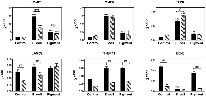
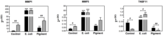
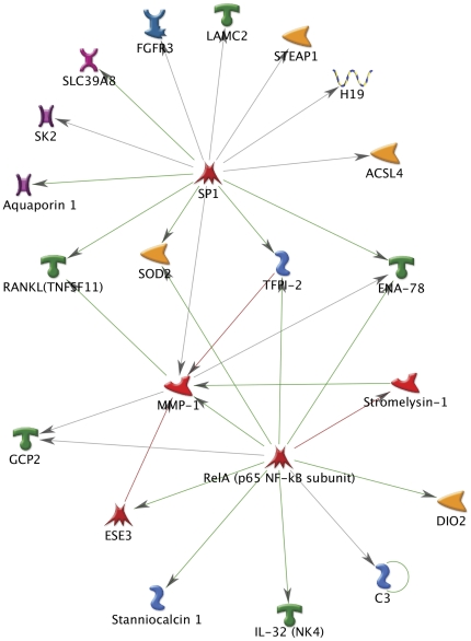
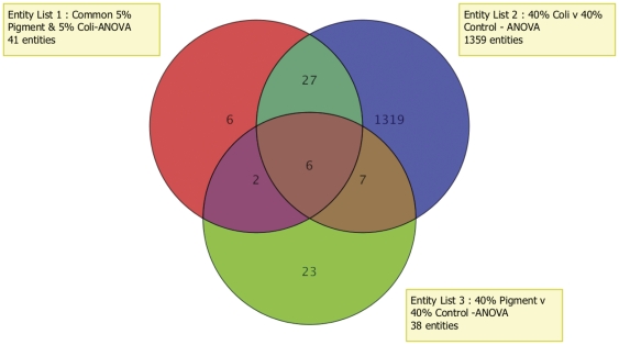
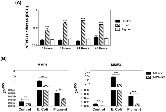
METHODOLOGY/PRINCIPAL FINDINGS: Gene expression profile analysis of human TM cells phagocytically challenged to E. coli or pigment under physiological and oxidative stress environment were performed using Affymetrix U133 plus 2.0 array and analyzed with Genespring GX. Despite the differential biological response elicited by E. coli and pigment particles, a number of genes, including MMP1, MMP3, TNFSF11, DIO2, KYNU, and KCCN2 showed differential expression with both phagocytic ligands in all conditions. Data was confirmed by qPCR in both human and porcine TM cells. Metacore pathway analysis and the usage of recombinant adenovirus encoding the dominant negative mutant of IkB identified NF-κB as a transcription factor mediating the up-regulation of at least MMP1 and MMP3 in TM cells with phagocytosis. In-gel zymography demonstrated increased collagenolytic and caseinolytic activities in the culture media of TM cells challenge to E. coli. In addition, collagenolytic I activity was further confirmed using the self-quenched fluorescent substrate DQ-Collagen I.
CONCLUSIONS/SIGNIFICANCE: Here we report for the first time the differential gene expression profile of TM cells phagocytically challenged with either E. coli or pigment. Our data indicate a potential role of phagocytosis in outflow pathway tissue homeostasis through the up-regulation and/or proteolytic activation of extracellular matrix remodeling genes.
小梁网(TM)中的细胞负责将房水从眼睛中排出,已知其具有高度吞噬作用。TM 细胞的吞噬功能被认为在流出通路的正常功能中发挥着重要作用。吞噬作用的功能障碍可能导致流出阻力异常和眼内压(IOP)升高。然而,TM 细胞吞噬作用引发的分子机制尚完全不清楚。
方法/主要发现:在生理和氧化应激环境下,用 Affymetrix U133 plus 2.0 阵列对人 TM 细胞进行了吞噬大肠杆菌或色素颗粒的基因表达谱分析,并使用 Genespring GX 进行了分析。尽管大肠杆菌和色素颗粒引起的生物学反应不同,但包括 MMP1、MMP3、TNFSF11、DIO2、KYNU 和 KCCN2 在内的许多基因在所有条件下均显示出与两种吞噬配体的差异表达。在人和猪 TM 细胞中通过 qPCR 进行了数据验证。Metacore 通路分析和使用编码 IkB 显性负突变体的重组腺病毒,鉴定了 NF-κB 作为介导 TM 细胞吞噬作用中 MMP1 和 MMP3 上调的转录因子。胶内酶谱分析表明,大肠杆菌刺激 TM 细胞培养物中胶原酶和酪蛋白酶活性增加。此外,使用自猝灭荧光底物 DQ-Collagen I 进一步证实了胶原酶 I 活性。
结论/意义:我们首次报道了 TM 细胞吞噬大肠杆菌或色素颗粒后差异的基因表达谱。我们的数据表明吞噬作用通过上调和/或细胞外基质重塑基因的蛋白水解激活,在流出通路组织稳态中具有潜在作用。