Mahdy Mohamed Ae Soliman, Eid Mohamed Z, Mohammed Mahmoud Abdel-Badei, Hafez Amr, Bhatia Jagdish
Ophthalmic Department, Al-Hussein University Hospital, Al-Azhar University, Cairo, Egypt.
Clin Ophthalmol. 2012;6:503-10. doi: 10.2147/OPTH.S29865. Epub 2012 Mar 29.
To assess the relationship between postoperative endothelial cell loss and microcoaxial phaco parameters using Ozil IP (Alcon Laboratories, Inc, Fort Worth, TX) in noncomplicated cataract surgery.
In this prospective observational study, 120 consecutive cases of cataract patients with different grades of nuclear hardness underwent microcoaxial phacoemulsification through a 2.2-mm clear corneal incision. An Alcon Infinity Vision System with Ozil IP (Alcon Laboratories) was used with an Ozil torsional handpiece and a Kelman-style 45° phacoemulsification tip. Patients underwent preoperative and postoperative central endothelial cell counts.
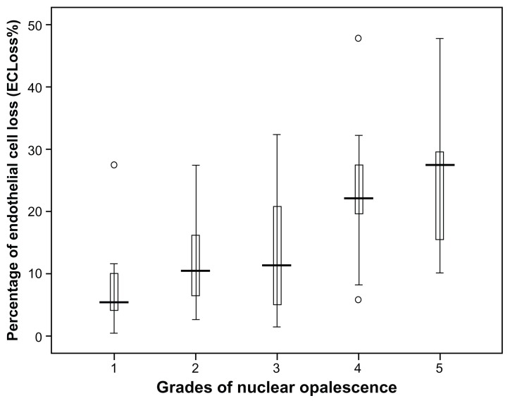
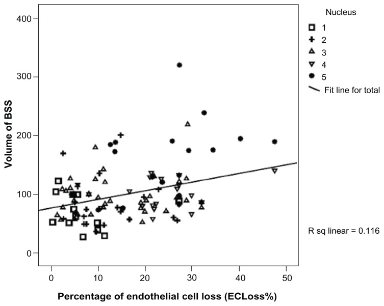
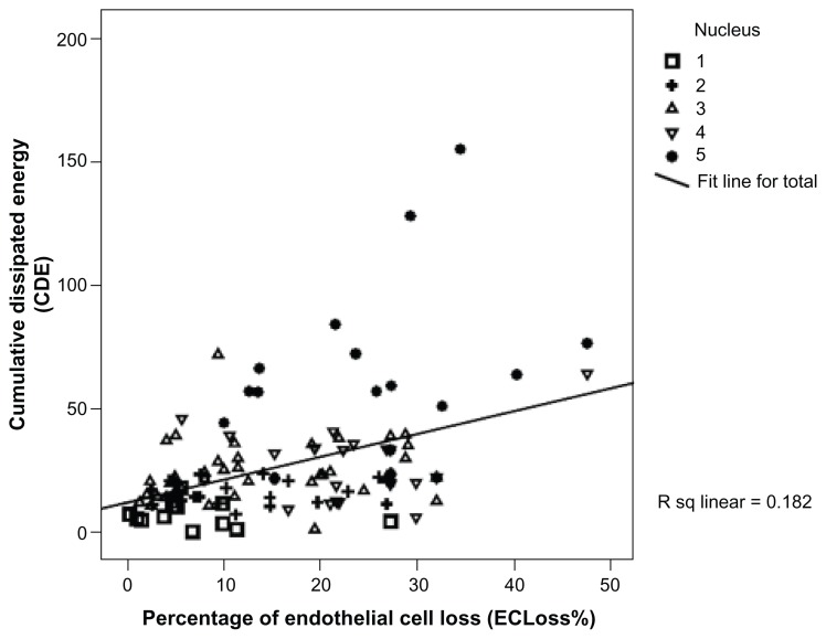
The study included 120 cases of age-related cataract whose mean age (standard deviation [SD]) was 59.68 years (9.47). There was a highly statistically significant endothelial cell loss (P < 0.001). The endothelial cell loss ranged 11-1149 cells/mm(2) with a median (interquartile range) of 386 cells/mm(2) (184.5-686 cells/mm(2)). The percentage of postoperative ECLoss% ranged from 0.48% to 47.8% with a median (interquartile range) of 15.4% (7.2% to 26.8%). A significant positive correlation was found between the ECLoss% and different phaco parameters. The Spearman's rank-order correlation coefficient values, rho, (ρ) were as follows: CDE (ρ = 0.425), aspiration time (ρ = 0.176), and volume (ρ = 0.278). Also, ECLoss% was significantly correlated with the grade of nuclear opalescence (Kendall's tau τ = 0.42).
Microcoaxial phacoemulsification was efficient in removing noncomplicated cataracts; however a statistically significant endothelial cell loss was noted, especially with increased nuclear hardness. This endothelial cell loss was mostly related to the increased cumulative dissipated energy (CDE), aspiration time, and volume of balanced salt solution used.
在非复杂性白内障手术中,使用奥兹尔IP(爱尔康实验室公司,沃思堡,德克萨斯州)评估术后内皮细胞丢失与微同轴超声乳化参数之间的关系。
在这项前瞻性观察研究中,120例连续的不同核硬度等级的白内障患者通过2.2毫米透明角膜切口进行微同轴超声乳化术。使用配备奥兹尔IP(爱尔康实验室)的爱尔康Infinity视觉系统,带有奥兹尔扭转式手持件和凯尔曼式45°超声乳化针头。患者术前和术后进行中央内皮细胞计数。
该研究纳入120例年龄相关性白内障患者,平均年龄(标准差[SD])为59.68岁(9.47)。存在高度统计学意义的内皮细胞丢失(P < 0.001)。内皮细胞丢失范围为11 - 1149个细胞/mm²,中位数(四分位间距)为386个细胞/mm²(184.5 - 686个细胞/mm²)。术后内皮细胞丢失百分比(ECLoss%)范围为0.48%至47.8%,中位数(四分位间距)为15.4%(7.2%至26.8%)。发现ECLoss%与不同的超声乳化参数之间存在显著正相关。斯皮尔曼等级相关系数值,rho(ρ)如下:累积消散能量(CDE)(ρ = 0.425)、抽吸时间(ρ = 0.176)和液体量(ρ = 0.278)。此外,ECLoss%与核混浊等级显著相关(肯德尔tau系数τ = 0.42)。
微同轴超声乳化术在去除非复杂性白内障方面有效;然而,注意到存在统计学意义的内皮细胞丢失,尤其是随着核硬度增加。这种内皮细胞丢失主要与累积消散能量(CDE)增加、抽吸时间以及平衡盐溶液使用量有关。