CAS Key Laboratory for Biological Effects of Nanomaterials and Nanosafety, National Center for Nanoscience and Technology, No. 11, First North Road, Zhongguancun, Beijing, China 100190.
ACS Nano. 2012 May 22;6(5):4483-93. doi: 10.1021/nn301282m. Epub 2012 May 4.
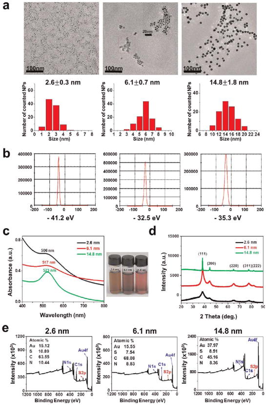
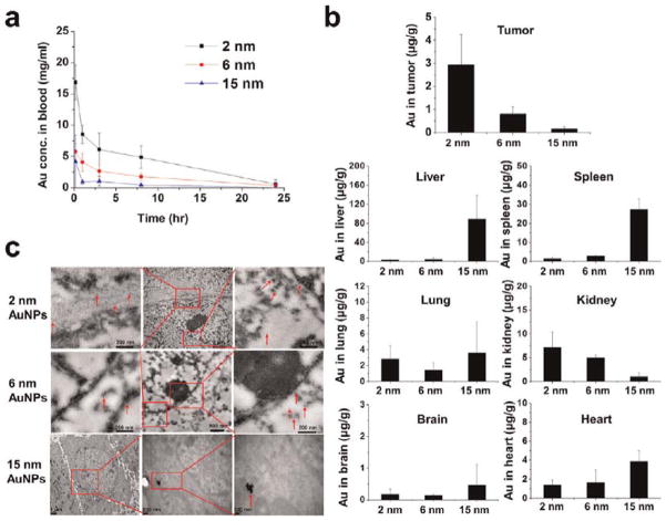
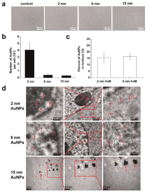
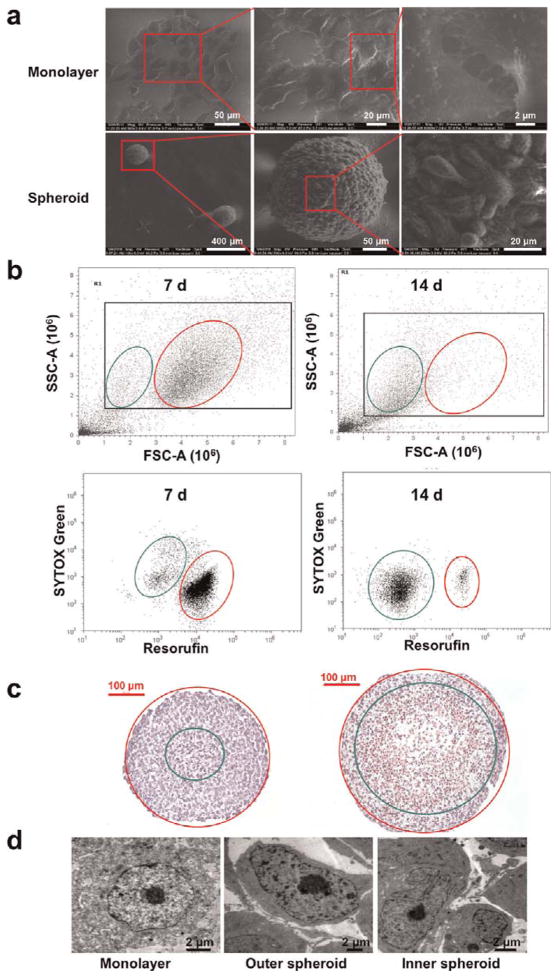
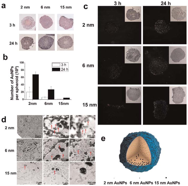
This work demonstrated that ultrasmall gold nanoparticles (AuNPs) smaller than 10 nm display unique advantages over nanoparticles larger than 10 nm in terms of localization to, and penetration of, breast cancer cells, multicellular tumor spheroids, and tumors in mice. Au@tiopronin nanoparticles that have tunable sizes from 2 to 15 nm with identical surface coatings of tiopronin and charge were successfully prepared. For monolayer cells, the smaller the Au@tiopronin NPs, the more AuNPs found in each cell. In addition, the accumulation of Au NPs in the ex vivo tumor model was size-dependent: smaller AuNPs were able to penetrate deeply into tumor spheroids, whereas 15 nm nanoparticles were not. Owing to their ultrasmall nanostructure, 2 and 6 nm nanoparticles showed high levels of accumulation in tumor tissue in mice after a single intravenous injection. Surprisingly, both 2 and 6 nm Au@tiopronin nanoparticles were distributed throughout the cytoplasm and nucleus of cancer cells in vitro and in vivo, whereas 15 nm Au@tiopronin nanoparticles were found only in the cytoplasm, where they formed aggregates. The ex vivo multicellular spheroid proved to be a good model to simulate in vivo tumor tissue and evaluate nanoparticle penetration behavior. This work gives important insights into the design and functionalization of nanoparticles to achieve high levels of accumulation in tumors.
这项工作表明,在定位和穿透乳腺癌细胞、多细胞肿瘤球体和小鼠肿瘤方面,小于 10nm 的超小金纳米颗粒(AuNPs)比大于 10nm 的纳米颗粒具有独特的优势。成功制备了具有从 2nm 到 15nm 可调尺寸的 Au@tiopronin 纳米颗粒,且具有相同的 tiopronin 表面涂层和电荷。对于单层细胞,Au@tiopronin NPs 越小,每个细胞中发现的 AuNPs 就越多。此外,Au NPs 在离体肿瘤模型中的积累具有尺寸依赖性:较小的 AuNPs 能够深入穿透肿瘤球体,而 15nm 纳米颗粒则不能。由于其超小的纳米结构,在单次静脉注射后,2nm 和 6nm 纳米颗粒在小鼠肿瘤组织中表现出高积累水平。令人惊讶的是,2nm 和 6nm 的 Au@tiopronin 纳米颗粒在体外和体内均分布于癌细胞的细胞质和细胞核中,而 15nm 的 Au@tiopronin 纳米颗粒仅分布于细胞质中,在细胞质中形成聚集体。离体多细胞球体被证明是模拟体内肿瘤组织和评估纳米颗粒穿透行为的良好模型。这项工作为设计和功能化纳米颗粒以实现肿瘤内高积累提供了重要的见解。