Fukuda Masamichi, Sasaki Hiroshi
Department of Ophthalmology, Kanazawa Medical University, Uchinada, Japan.
Clin Ophthalmol. 2012;6:585-93. doi: 10.2147/OPTH.S30935. Epub 2012 Apr 17.
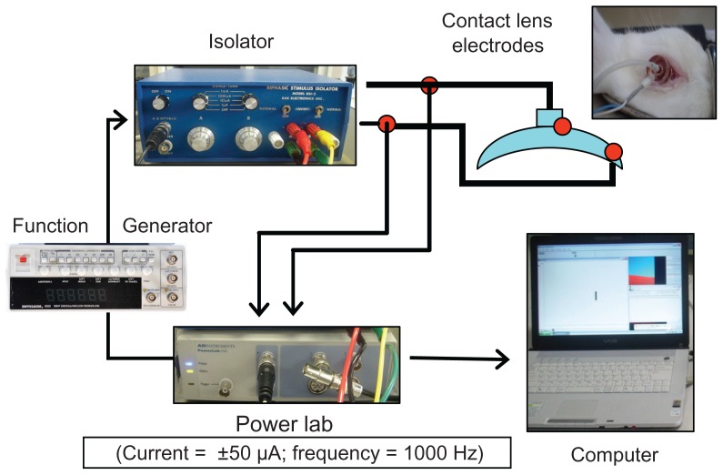
We attempted to develop a device for measuring electrical corneal resistance (CR) using corneal contact lens electrodes to quantitatively evaluate corneal injury in vivo. In the present study, full-thickness detachment of the corneal epithelium was induced by n-heptanol, and the feasibility of the quantitative evaluation of this injury by corneal contact lens electrodes was evaluated in vivo.
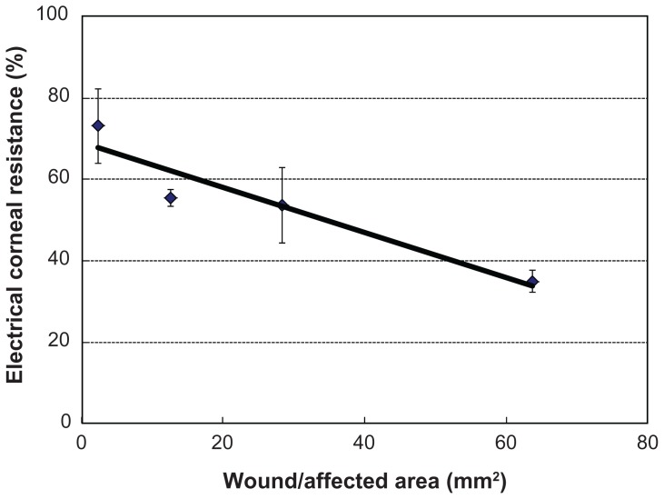
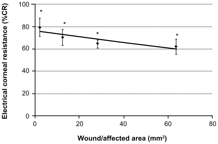
The central area of an albino rabbit's cornea was exposed to a filter paper pre-immersed in n-heptanol for 1 minute to induce injury of the corneal epithelium. After induction of injury, the electrical CR was measured and the percentage of CR (%CR) was calculated. Fluorescein solution (3 μL) was applied to the wound/affected area of the corneal epithelium for photography with a slit-lamp biomicroscope. The wound/affected area was measured using an image analysis system. The correlation between the %CR and the wound/affected area was analyzed.
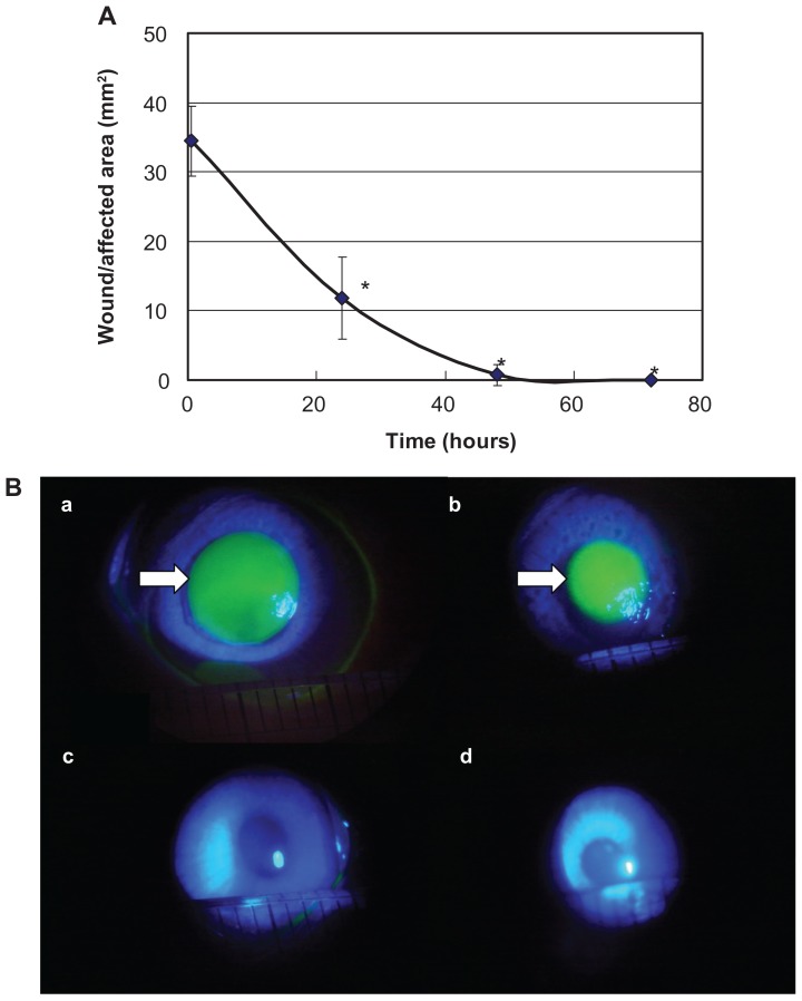
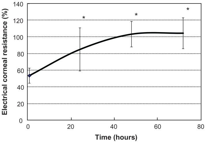
As the size of the wound/affected area of the corneal epithelium increased, the %CR decreased after corneal epithelium detachment. Thus, a close correlation was found between the area of corneal epithelium detachment and the %CR.
The corneal resistance device that we developed was capable of quantitatively evaluating n-heptanol-induced full-thickness injuries of the corneal epithelium.
我们试图开发一种使用角膜接触镜电极测量角膜电阻(CR)的装置,以在体内定量评估角膜损伤。在本研究中,用正庚醇诱导角膜上皮全层脱离,并在体内评估角膜接触镜电极对这种损伤进行定量评估的可行性。
将白化兔角膜的中央区域暴露于预先浸入正庚醇的滤纸上1分钟,以诱导角膜上皮损伤。诱导损伤后,测量电CR并计算CR百分比(%CR)。将荧光素溶液(3μL)应用于角膜上皮的伤口/受影响区域,用裂隙灯生物显微镜进行拍照。使用图像分析系统测量伤口/受影响区域。分析%CR与伤口/受影响区域之间的相关性。
随着角膜上皮伤口/受影响区域的大小增加,角膜上皮脱离后%CR降低。因此,发现角膜上皮脱离面积与%CR之间存在密切相关性。
我们开发的角膜电阻装置能够定量评估正庚醇诱导的角膜上皮全层损伤。