Department of Diagnostic Radiology, Chonnam National University Medical School, Chonnam National University Hwasun Hospital, Hwasun 519-763, Korea.
Korean J Radiol. 2012 Jan-Feb;13 Suppl 1(Suppl 1):S89-97. doi: 10.3348/kjr.2012.13.S1.S89. Epub 2012 Apr 23.
The purpose of this study was to prospectively compare pre-operative computed tomography (CT) perfusion parameters with tumor grade from colorectal adenocarcinoma (CRC) and to correlate pre-operative CT perfusion parameters with microvessel density (MVD) to evaluate angiogenesis in CRC.
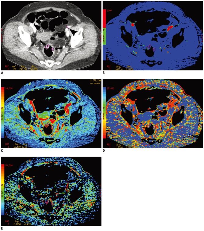
Pre-operative perfusion CTs were performed with a 64-channel multidetector row CT in 27 patients (17 women and 10 men; age range 32-82 years) who were diagnosed with CRC involving the sigmoid and rectum between August 2006 and November 2007. All patients underwent surgery without pre-operative chemotherapy or radiation therapy. Dynamic perfusion CTs were performed for 65 seconds after intravenous injection of contrast medium (100 mL, 300 mg of iodine per mL, 5 mL/sec). Before surgery, blood flow (BF), blood volume, mean transit time (MTT), and permeability-surface area product were measured in the tumor. After surgery, one gastrointestinal pathologist evaluated tumor grade and performed immunohistochemical staining using CD 34 to determine MVD in each tumor. The Kruskal-Wallis test was used to compare CT perfusion parameters with tumor grade, and Pearson's correlation analysis was used to correlate CT perfusion parameters with MVD.
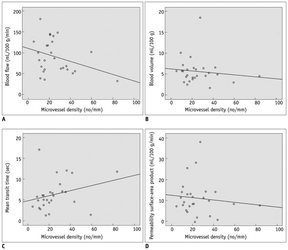
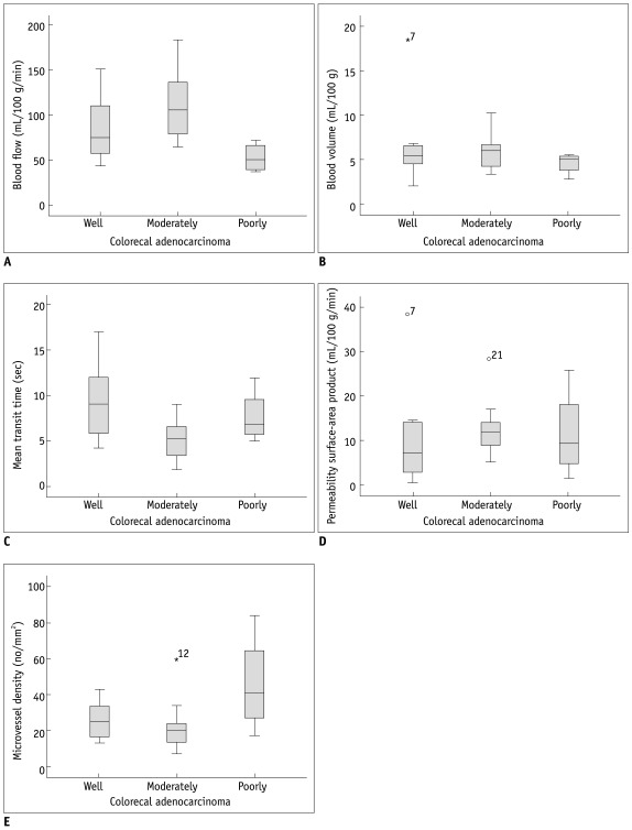
In 27 patients with CRC, tumor grading was as follows: well differentiated (n = 8); moderately differentiated (n = 15); and poorly differentiated (n = 4). BF was higher in moderately differentiated CRC than well differentiated and poorly differentiated CRCs (p = 0.14). MTT was shorter in moderately differentiated than well differentiated and poorly differentiated CRCs (p = 0.039). The MVD was greater in poorly differentiated than well differentiated and moderately differentiated CRCs (p = 0.034). There was no significant correlation between other perfusion parameters and tumor grade. There was no significant correlation between CT perfusion parameters and MVD.
BF and MTT measurement by perfusion CT is effective in predicting moderately differentiated CRCs. However, perfusion CT is limited in distinguishing well differentiated and poorly differentiated CRCs. Pre-operative perfusion CT does not reflect the MVD of CRCs.
本研究旨在前瞻性比较结直肠腺癌(CRC)术前 CT 灌注参数与肿瘤分级,并将术前 CT 灌注参数与微血管密度(MVD)相关联,以评估 CRC 中的血管生成。
2006 年 8 月至 2007 年 11 月,对 27 例诊断为累及乙状结肠和直肠的 CRC 患者(17 名女性和 10 名男性;年龄 32-82 岁)进行了术前灌注 CT 检查。所有患者均未接受术前化疗或放疗即行手术治疗。静脉注射造影剂(100mL,300mg 碘/mL,5mL/sec)后进行 65 秒动态灌注 CT 检查。术前,测量肿瘤内的血流量(BF)、血容量、平均通过时间(MTT)和通透性表面积乘积。术后,一名胃肠病理学家评估肿瘤分级,并使用 CD34 进行免疫组织化学染色以确定每个肿瘤的 MVD。采用 Kruskal-Wallis 检验比较 CT 灌注参数与肿瘤分级,采用 Pearson 相关分析比较 CT 灌注参数与 MVD。
在 27 例 CRC 患者中,肿瘤分级如下:高分化(n=8);中分化(n=15);低分化(n=4)。中分化 CRC 的 BF 高于高分化和低分化 CRC(p=0.14)。中分化 CRC 的 MTT 短于高分化和低分化 CRC(p=0.039)。低分化 CRC 的 MVD 大于高分化和中分化 CRC(p=0.034)。其他灌注参数与肿瘤分级之间无显著相关性。CT 灌注参数与 MVD 之间无显著相关性。
灌注 CT 测量 BF 和 MTT 可有效预测中分化 CRC。然而,灌注 CT 难以区分高分化和低分化 CRC。术前灌注 CT 不能反映 CRC 的 MVD。