Ocular Surface Imaging Center, Cornea Service, Massachusetts Eye & Ear Infirmary, Department of Ophthalmology, Harvard Medical School, Boston, Massachusetts 02114, USA.
Ophthalmology. 2012 Sep;119(9):1791-7. doi: 10.1016/j.ophtha.2012.03.005. Epub 2012 May 16.
To analyze the morphologic features of corneal epithelial cells and keratocytes by in vivo confocal microscopy in patients with herpes simplex keratitis (HSK) as associated with corneal innervation.
Prospective, cross-sectional, controlled, single-center study.
Thirty-one eyes with the diagnosis HSK and their contralateral clinically unaffected eyes were studied and compared with normal controls (n = 15).
In vivo confocal microscopy (Confoscan 4; Nidek Technologies, Gamagori, Japan) and corneal esthesiometry (Cochet-Bonnet; Luneau Ophthalmologie, Chartres, France) of the central cornea were performed bilaterally in all patients and controls. Patients were grouped into normal (>5.5 cm), mild (>2.5-5.5 cm), and severe (<2.5 cm) loss of sensation.
Changes in morphologic features and density of the superficial and basal epithelial cells, as well as stromal keratocytes, were assessed by 2 masked observers. Changes were correlated to corneal sensation, number of nerves, and total length of nerves.
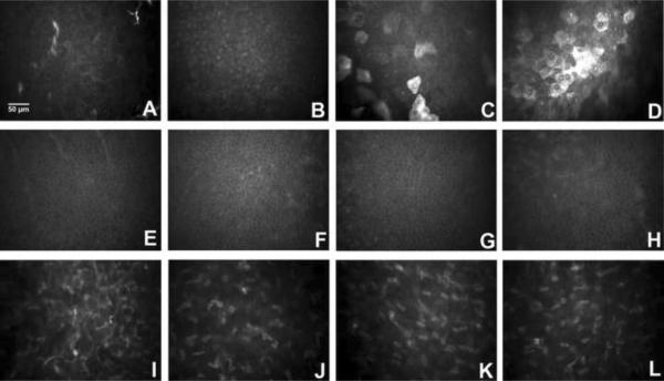
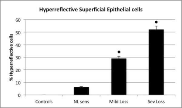
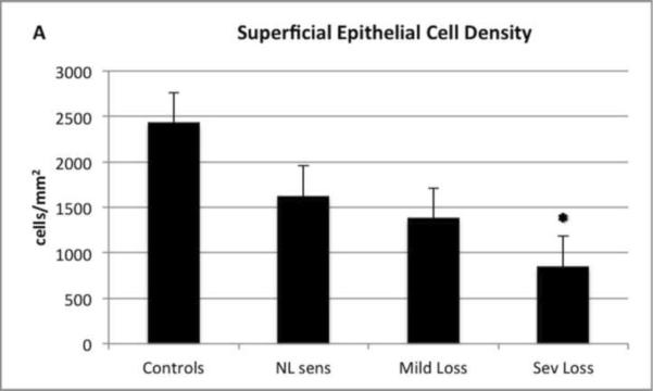
There was a significant and gradual decrease in the density of superficial epithelial cells in HSK eyes, with 852.50 ± 24.4 cells/mm(2) in eyes with severe sensation loss and 2435.23 ± 224.3 cells/mm(2) in control eyes (P = 0.008). Superficial epithelial cell size was 2.5-fold larger in HSK eyes (835.3 μm(2)) compared with contralateral or normal eyes (407.4 μm(2); P = 0.003). A significant number of hyperreflective desquamating superficial epithelial cells were present in HSK eyes with normal (6.4%), mild (29.1%), and severe (52.2%) loss of sensation, but were absent in controls. The density of basal epithelial cells, anterior keratocytes, and posterior keratocytes did not show statistical significance between patients and controls. Changes in superficial epithelial cell density and morphologic features correlated strongly with total nerve length, number, and corneal sensation. Scans of contralateral eyes did not show any significant epithelial or stromal changes compared with controls.
In vivo confocal microscopy reveals profound HSK-induced changes in the superficial epithelium, as demonstrated by increase in cell size, decrease in cell density, and squamous metaplasia. This study demonstrated that these changes correlate strongly with changes in corneal innervation.
通过共聚焦显微镜对单纯疱疹性角膜炎(HSK)患者角膜神经支配相关的角膜上皮细胞和角膜基质细胞的形态学特征进行分析。
前瞻性、横断面、对照、单中心研究。
共纳入 31 只诊断为 HSK 的患眼及其对侧临床未受影响的眼,并与正常对照组(n=15)进行比较。
所有患者和对照组均行双侧中央角膜共聚焦显微镜(Confoscan 4;日本尼德克技术公司)和角膜知觉检查(Cochet-Bonnet;法国 Luneau 眼科公司)。根据角膜感觉的丧失程度,将患者分为正常(>5.5cm)、轻度(>2.5-5.5cm)和重度(<2.5cm)。
由 2 名盲法观察者评估上皮细胞和基质基质细胞的超微结构和密度的变化。这些变化与角膜感觉、神经数量和神经总长度相关联。
HSK 眼的表层上皮细胞密度呈显著且逐渐下降,严重感觉丧失眼的密度为 852.50±24.4 个/平方毫米,而正常对照组的密度为 2435.23±224.3 个/平方毫米(P=0.008)。HSK 眼的表层上皮细胞大小是对侧或正常眼的 2.5 倍(835.3μm2)(P=0.003)。在感觉正常(6.4%)、轻度(29.1%)和重度(52.2%)丧失的 HSK 眼中,均可见到大量的超反射性脱屑的表层上皮细胞,但在对照组中未见。HSK 患者与对照组之间,基底上皮细胞、前基质细胞和后基质细胞的密度无统计学差异。表层上皮细胞密度和形态学特征的变化与神经总长度、数量和角膜感觉强烈相关。与对照组相比,对侧眼的扫描未见明显的上皮或基质变化。
共聚焦显微镜显示 HSK 可导致明显的表层上皮改变,表现为细胞大小增加、细胞密度降低和鳞状化生。本研究表明这些变化与角膜神经支配的变化密切相关。