Departments of Neurology (X.Z., H.M., M.M.W.), Molecular and Integrative, Physiology (J.S., M.M.W.), and Pathology (M.B.), University of Michigan Medical School, Ann Arbor, MI 48109; Department of Neurology, Veterans Administration Ann Arbor Healthcare, System, Ann Arbor, MI 48105 (M.M.W); Institute for Neuropathology, UniversitatsSpital, CH-8091 Zurich (E.J.R.); Department of Pathology and Center for Alzheimer's Disease and Related Disorders, Southern Illinois University School of Medicine, Springfield, IL 62794 (B.E.M.); Departments of Pathology (M. B.S.L.), Neurology (B.B.W.), and Public Health Sciences (B.B.W.), University of Virginia, Charlottesville, VA 22908.
Transl Stroke Res. 2012 Mar;3(1):138-45. doi: 10.1007/s12975-011-0112-2. Epub 2011 Oct 20.
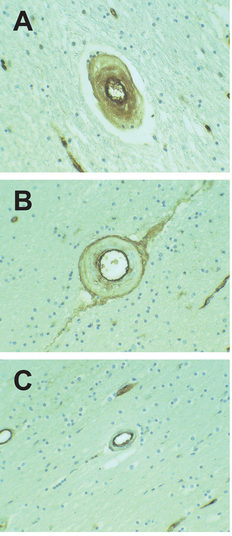
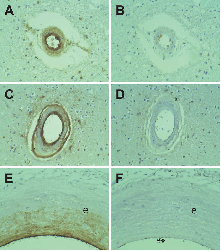
CADASIL (cerebral autosomal dominant arteriopathy subcortical infarcts and leukoencephalopathy) is a genetic disorder hallmarked by ischemic stroke and vascular dementia. Characteristic pathological changes in the vasculature include thickening of small arteries and accumulation of heterogeneous material within the vessel wall. We tested whether endothelial von Willebrand factor (vWF) accumulates in CADASIL vessels and whether exposure of smooth muscle cells to vWF alters the expression of smooth muscle gene expression.
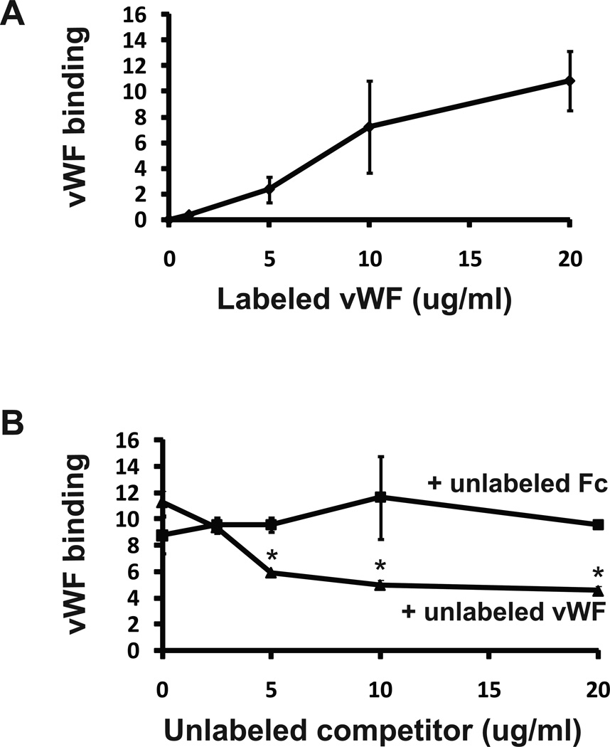
Brain sections obtained at autopsy from six North American CADASIL patients were examined using immunohistochemistry for vWF and IgG. Rat aortic smooth muscle cells (A7R5 cells) were tested for binding to infrared-tag labeled vWF. Finally, A7R5 cells were exposed to vWF, and expression of mature smooth muscle marker genes was analyzed by quantitative reverse transcriptase PCR.
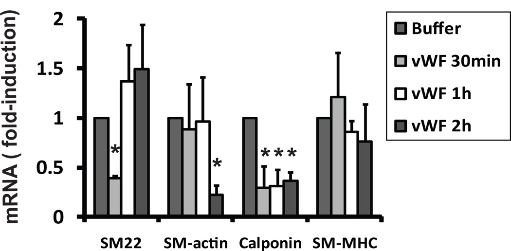
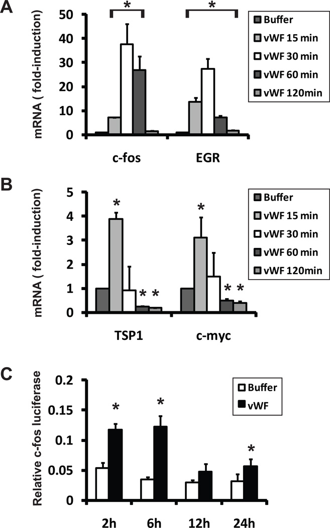
vWF is expressed in the penetrating arterial walls in all CADASIL samples. IgG, a marker of serum extravasation, was present only in a minority of arterial walls. vWF binds to smooth muscle cells in vitro, and low concentrations of vWF rapidly activate c-fos, EGR, TSP1, and c-myc while specifically inhibiting RNA encoding smooth muscle actin, calponin, and SM22.
These data demonstrate that vWF, likely produced by the endothelium, permeates the vessel wall of CADASIL brains. Exposure of smooth muscle cells to vWF results in reduction of specific RNAs required for normal vascular homeostasis. This is the first report of accumulation of a protein within CADASIL vessels that inhibits vascular gene expression and implicates a role for vWF beyond hemostasis.
CADASIL(伴有皮质下梗死和白质脑病的常染色体显性遗传性脑动脉病)是一种遗传疾病,以缺血性中风和血管性痴呆为特征。血管的特征性病理变化包括小动脉增厚和血管壁内异质物质的积累。我们检测了 CADASIL 血管中内皮 von Willebrand 因子(vWF)是否积累,以及平滑肌细胞暴露于 vWF 是否改变平滑肌基因表达。
使用免疫组织化学方法检测 6 名北美 CADASIL 患者尸检脑组织切片中的 vWF 和 IgG。用红外标记的 vWF 检测大鼠主动脉平滑肌细胞(A7R5 细胞)的结合情况。最后,将 A7R5 细胞暴露于 vWF,通过定量逆转录 PCR 分析成熟平滑肌标志物基因的表达。
vWF 在所有 CADASIL 样本的穿透性动脉壁中表达。IgG,一种血清外渗的标志物,仅存在于少数动脉壁中。vWF 在体外与平滑肌细胞结合,低浓度的 vWF 可迅速激活 c-fos、EGR、TSP1 和 c-myc,同时特异性抑制编码平滑肌肌动蛋白、钙调蛋白和 SM22 的 RNA。
这些数据表明,vWF 可能由内皮细胞产生,穿透 CADASIL 大脑血管壁。平滑肌细胞暴露于 vWF 导致正常血管稳态所需的特定 RNA 减少。这是 CADASIL 血管中积累一种抑制血管基因表达的蛋白质的首次报道,并暗示 vWF 在止血之外发挥作用。