Laboratory for Cell Signaling, RIKEN Research Center for Allergy and Immunology, Yokohama, Kanagawa 230-0045, Japan.
J Exp Med. 2012 Jun 4;209(6):1201-17. doi: 10.1084/jem.20112741. Epub 2012 May 28.
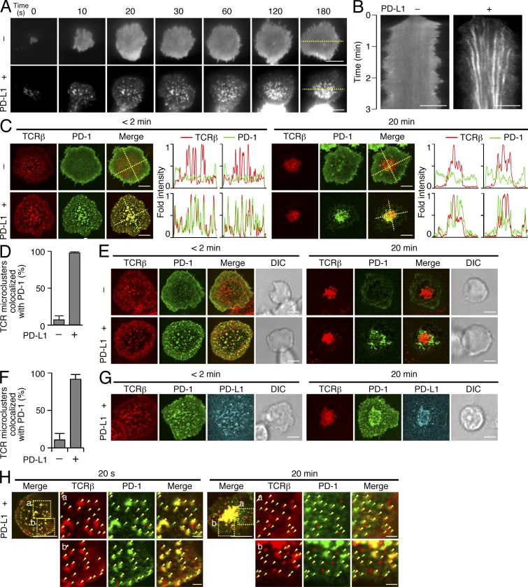
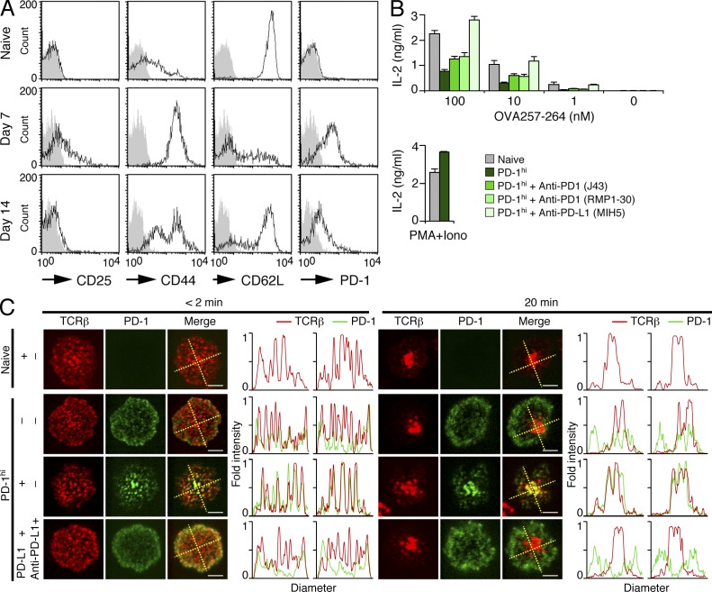
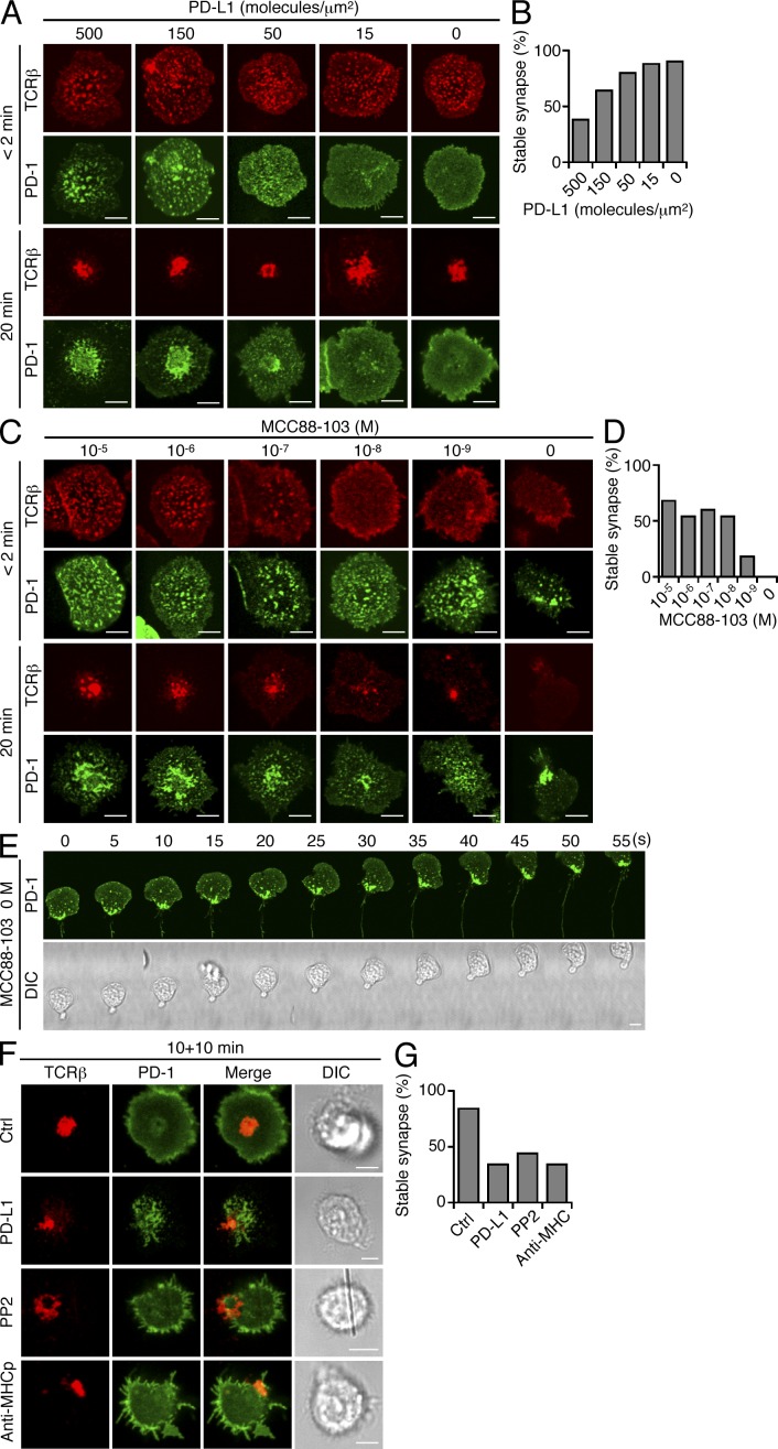
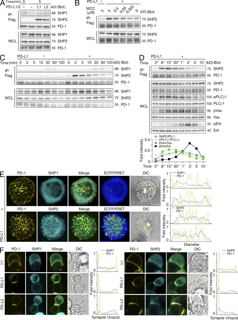
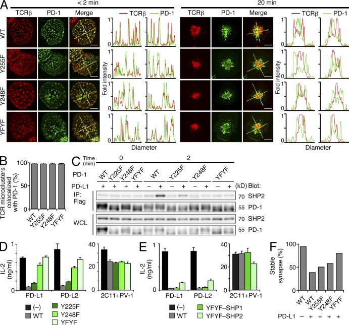
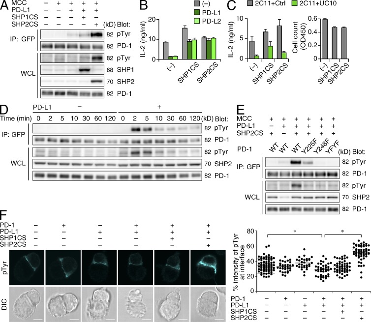
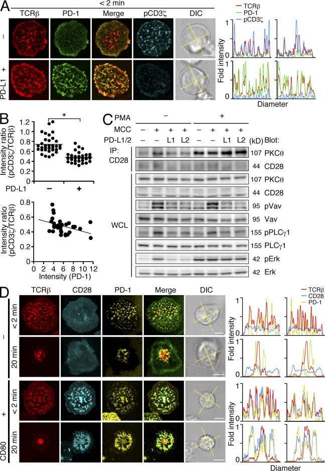
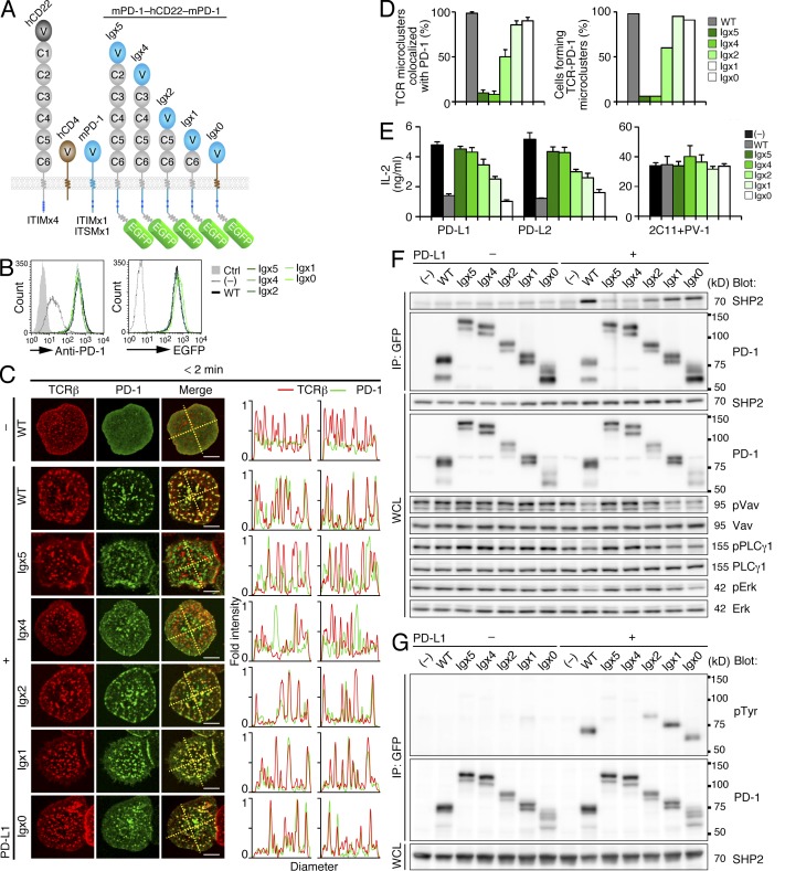
Programmed cell death 1 (PD-1) is a negative costimulatory receptor critical for the suppression of T cell activation in vitro and in vivo. Single cell imaging elucidated a molecular mechanism of PD-1-mediated suppression. PD-1 becomes clustered with T cell receptors (TCRs) upon binding to its ligand PD-L1 and is transiently associated with the phosphatase SHP2 (Src homology 2 domain-containing tyrosine phosphatase 2). These negative costimulatory microclusters induce the dephosphorylation of the proximal TCR signaling molecules. This results in the suppression of T cell activation and blockade of the TCR-induced stop signal. In addition to PD-1 clustering, PD-1-TCR colocalization within microclusters is required for efficient PD-1-mediated suppression. This inhibitory mechanism also functions in PD-1(hi) T cells generated in vivo and can be overridden by a neutralizing anti-PD-L1 antibody. Therefore, PD-1 microcluster formation is important for regulation of T cell activation.
程序性细胞死亡蛋白 1(PD-1)是一种负性共刺激受体,对于体外和体内 T 细胞的激活具有抑制作用。单细胞成像揭示了 PD-1 介导的抑制的分子机制。PD-1 在与配体 PD-L1 结合后与 T 细胞受体(TCRs)聚集,并与磷酸酶 SHP2(Src 同源 2 结构域酪氨酸磷酸酶 2)短暂相关。这些负性共刺激微簇诱导 TCR 信号分子的去磷酸化。这导致 T 细胞激活的抑制和 TCR 诱导的停止信号的阻断。除了 PD-1 聚集外,微簇内的 PD-1-TCR 共定位对于有效的 PD-1 介导的抑制也是必需的。这种抑制机制也在体内产生的 PD-1(hi)T 细胞中起作用,并且可以被中和抗 PD-L1 抗体所克服。因此,PD-1 微簇的形成对于 T 细胞激活的调节很重要。