• 文献检索
  • 文档翻译
  • 深度研究
  • 学术资讯
  • Suppr Zotero 插件Zotero 插件
  • 邀请有礼
  • 套餐&价格
  • 历史记录
应用&插件
Suppr Zotero 插件Zotero 插件浏览器插件Mac 客户端Windows 客户端微信小程序
定价
高级版会员购买积分包购买API积分包
服务
文献检索文档翻译深度研究API 文档MCP 服务
关于我们
关于 Suppr公司介绍联系我们用户协议隐私条款
关注我们

Suppr 超能文献

核心技术专利:CN118964589B侵权必究
粤ICP备2023148730 号-1Suppr @ 2026

文献检索

告别复杂PubMed语法,用中文像聊天一样搜索,搜遍4000万医学文献。AI智能推荐,让科研检索更轻松。

立即免费搜索

文件翻译

保留排版,准确专业,支持PDF/Word/PPT等文件格式,支持 12+语言互译。

免费翻译文档

深度研究

AI帮你快速写综述,25分钟生成高质量综述,智能提取关键信息,辅助科研写作。

立即免费体验

颅内 EEG 表面重建:正常和异常大脑功能的新见解。

Intracranial EEG surface renderings: new insights into normal and abnormal brain function.

机构信息

Department of Neurology, Yale University School of Medicine, New Haven, CT 06520-8018, USA.

出版信息

Neuroscientist. 2013 Jun;19(3):238-47. doi: 10.1177/1073858412447876. Epub 2012 May 31.

DOI:10.1177/1073858412447876
PMID:22653695
原文链接:https://pmc.ncbi.nlm.nih.gov/articles/PMC6343473/
Abstract

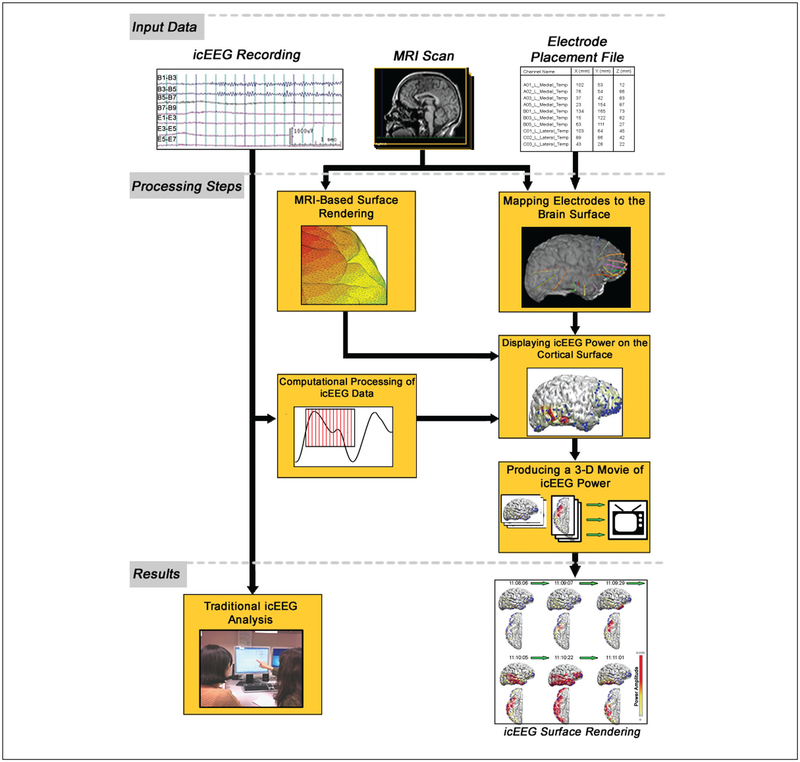
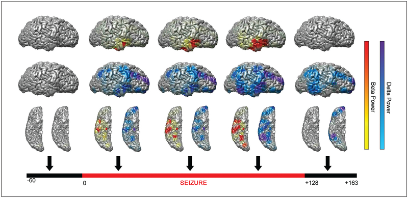
Intracranial electro-encephalography (icEEG) provides a unique opportunity to record directly from the human brain and is clinically important for planning epilepsy surgery. However, traditional visual analysis of icEEG is often challenging. The typical simultaneous display of multiple electrode channels can prevent an in-depth understanding of the spatial-time course of brain activity. In recent decades, advances in the field of neuroimaging have provided powerful new tools for the analysis and display of signals in the brain. These methods can now be applied to icEEG to map electrical signal information onto a three-dimensional rendering of a patient's cortex and graphically observe the changes in voltage over time. This approach provides rapid visualization of seizures and normal activity propagating over the brain surface and can also illustrate subtle changes that might be missed by traditional icEEG analysis. In addition, the direct mapping of signal information onto accurate anatomical structures can assist in the precise targeting of sites for epilepsy surgery and help correlate electrical activity with behavior. Bringing icEEG data into a standardized anatomical space will also enable neuroimaging methods of statistical analysis to be applied. As new technologies lead to a dramatic increase in the rate of data acquisition, these novel visualization and analysis techniques will play an important role in processing the valuable information obtained through icEEG.

摘要

颅内脑电图(icEEG)提供了直接记录人类大脑的独特机会,对规划癫痫手术具有重要的临床意义。然而,icEEG 的传统视觉分析通常具有挑战性。多个电极通道的典型同步显示可能会妨碍对大脑活动的时空过程的深入理解。近几十年来,神经影像学领域的进步为大脑信号的分析和显示提供了强大的新工具。这些方法现在可以应用于 icEEG,将电信号信息映射到患者皮质的三维渲染图上,并直观地观察电压随时间的变化。这种方法可以快速可视化脑表面传播的癫痫发作和正常活动,还可以说明传统 icEEG 分析可能会错过的细微变化。此外,将信号信息直接映射到准确的解剖结构上,可以帮助精确地确定癫痫手术的靶点,并有助于将电活动与行为相关联。将 icEEG 数据纳入标准化的解剖空间还将使神经影像学的统计分析方法得以应用。随着新技术导致数据采集速度的急剧增加,这些新颖的可视化和分析技术将在处理通过 icEEG 获得的有价值信息方面发挥重要作用。

https://cdn.ncbi.nlm.nih.gov/pmc/blobs/c2db/6343473/6e7d5d42740f/nihms-998558-f0004.jpg
https://cdn.ncbi.nlm.nih.gov/pmc/blobs/c2db/6343473/5a4b8ba29b69/nihms-998558-f0001.jpg
https://cdn.ncbi.nlm.nih.gov/pmc/blobs/c2db/6343473/8a051db97259/nihms-998558-f0002.jpg
https://cdn.ncbi.nlm.nih.gov/pmc/blobs/c2db/6343473/3d1be734726d/nihms-998558-f0003.jpg
https://cdn.ncbi.nlm.nih.gov/pmc/blobs/c2db/6343473/6e7d5d42740f/nihms-998558-f0004.jpg

相似文献

1
Intracranial EEG surface renderings: new insights into normal and abnormal brain function.颅内 EEG 表面重建:正常和异常大脑功能的新见解。
Neuroscientist. 2013 Jun;19(3):238-47. doi: 10.1177/1073858412447876. Epub 2012 May 31.
2
Seizure localization using three-dimensional surface projections of intracranial EEG power.使用颅内 EEG 功率的三维表面投影进行癫痫定位。
Neuroimage. 2013 Dec;83:616-26. doi: 10.1016/j.neuroimage.2013.07.010. Epub 2013 Jul 11.
3
Recording human electrocorticographic (ECoG) signals for neuroscientific research and real-time functional cortical mapping.记录用于神经科学研究和实时功能性皮层图谱绘制的人类皮层脑电图(ECoG)信号。
J Vis Exp. 2012 Jun 26(64):3993. doi: 10.3791/3993.
4
Magnetic source imaging and ictal SPECT in MRI-negative neocortical epilepsies: additional value and comparison with intracranial EEG.磁共振阴性局灶性皮质癫痫的磁源成像和发作期 SPECT:与颅内 EEG 的附加价值和比较。
Epilepsia. 2013 Feb;54(2):359-69. doi: 10.1111/epi.12004. Epub 2012 Oct 25.
5
Clinical significance of nonhabitual seizures during intracranial EEG monitoring.颅内 EEG 监测中非习惯性发作的临床意义。
Epilepsia. 2014 Jan;55(1):e1-5. doi: 10.1111/epi.12462. Epub 2013 Dec 2.
6
Evaluating the Safety of Simultaneous Intracranial Electroencephalography and Functional Magnetic Resonance Imaging Acquisition Using a 3 Tesla Magnetic Resonance Imaging Scanner.使用3特斯拉磁共振成像扫描仪评估同步颅内脑电图和功能磁共振成像采集的安全性。
Front Neurosci. 2022 Jun 23;16:921922. doi: 10.3389/fnins.2022.921922. eCollection 2022.
7
The correlation of magnetoencephalography to intracranial EEG in localizing the epileptogenic zone: a study of the surgical resection outcome.脑磁图与颅内脑电图在癫痫灶定位中的相关性:手术切除结果研究
Epilepsy Res. 2014 Nov;108(9):1581-90. doi: 10.1016/j.eplepsyres.2014.08.016. Epub 2014 Sep 3.
8
Seizure susceptibility and infraslow modulatory activity in the intracranial electroencephalogram.颅内脑电图中的发作易感性和亚慢波调制活动。
Epilepsia. 2018 Nov;59(11):2075-2085. doi: 10.1111/epi.14559. Epub 2018 Sep 6.
9
Neural Activity Elicited by a Cognitive Task can be Detected in Single-Trials with Simultaneous Intracerebral EEG-fMRI Recordings.通过同步颅内脑电图-功能磁共振成像记录,在单试验中可检测到由认知任务引发的神经活动。
Int J Neural Syst. 2017 Feb;27(1):1750001. doi: 10.1142/S0129065717500010. Epub 2016 Aug 22.
10
Accuracy of MEG in localizing irritative zone and seizure onset zone: Quantitative comparison between MEG and intracranial EEG.脑磁图在定位激惹区和癫痫发作起始区的准确性:脑磁图与颅内脑电图的定量比较
Epilepsy Res. 2016 Nov;127:291-301. doi: 10.1016/j.eplepsyres.2016.08.013. Epub 2016 Aug 16.

引用本文的文献

1
Influences of electrode density on intracranial seizure localisation: a single-blinded randomised crossover study.电极密度对颅内癫痫灶定位的影响:一项单盲随机交叉研究。
EBioMedicine. 2025 Mar;113:105606. doi: 10.1016/j.ebiom.2025.105606. Epub 2025 Mar 3.
2
Accuracy of omni-planar and surface casting of epileptiform activity for intracranial seizure localization.颅内癫痫灶定位中致痫样活动的全平面和表面描记的准确性。
Epilepsia. 2021 Apr;62(4):947-959. doi: 10.1111/epi.16841. Epub 2021 Feb 26.
3
Using 3D-Printed Mesh-Like Brain Cortex with Deep Structures for Planning Intracranial EEG Electrode Placement.

本文引用的文献

1
Improved Localizadon of Cortical Activity by Combining EEG and MEG with MRI Cortical Surface Reconstruction: A Linear Approach.通过将 EEG 和 MEG 与 MRI 皮质表面重建相结合来提高皮质活动的本地化:一种线性方法。
J Cogn Neurosci. 1993 Spring;5(2):162-76. doi: 10.1162/jocn.1993.5.2.162.
2
Deformable templates for face recognition.用于人脸识别的可变形模板。
J Cogn Neurosci. 1991 Winter;3(1):59-70. doi: 10.1162/jocn.1991.3.1.59.
3
Decoding covert spatial attention using electrocorticographic (ECoG) signals in humans.利用人类脑电皮层电图(ECoG)信号解码隐蔽的空间注意力。
使用带有深部结构的3D打印网状脑皮质进行颅内脑电图电极放置规划
J Digit Imaging. 2020 Apr;33(2):324-333. doi: 10.1007/s10278-019-00275-3.
4
Neural correlates of spatial and nonspatial attention determined using intracranial electroencephalographic signals in humans.利用人类颅内脑电图信号确定空间和非空间注意力的神经关联。
Hum Brain Mapp. 2016 Aug;37(8):3041-54. doi: 10.1002/hbm.23225. Epub 2016 Apr 29.
5
Seizure localization using three-dimensional surface projections of intracranial EEG power.使用颅内 EEG 功率的三维表面投影进行癫痫定位。
Neuroimage. 2013 Dec;83:616-26. doi: 10.1016/j.neuroimage.2013.07.010. Epub 2013 Jul 11.
Neuroimage. 2012 May 1;60(4):2285-93. doi: 10.1016/j.neuroimage.2012.02.017. Epub 2012 Feb 16.
4
Discrete gamma oscillations identify the seizure onset zone in some pediatric epilepsy patients.离散伽马振荡可识别部分小儿癫痫患者的癫痫发作起始区。
Annu Int Conf IEEE Eng Med Biol Soc. 2011;2011:3095-8. doi: 10.1109/IEMBS.2011.6090845.
5
Language mapping in multilingual patients: electrocorticography and cortical stimulation during naming.多语言患者的语言映射:命名过程中的皮质脑电图和皮质刺激
Front Hum Neurosci. 2011 Feb 22;5:13. doi: 10.3389/fnhum.2011.00013. eCollection 2011.
6
Tuning of the human neocortex to the temporal dynamics of attended events.人类新皮层对关注事件的时间动态的调整。
J Neurosci. 2011 Mar 2;31(9):3176-85. doi: 10.1523/JNEUROSCI.4518-10.2011.
7
3D visualization of subdural electrode shift as measured at craniotomy reopening.开颅术再切开时测量的硬膜下电极移位的 3D 可视化。
Epilepsy Res. 2011 Mar;94(1-2):102-9. doi: 10.1016/j.eplepsyres.2011.01.011. Epub 2011 Feb 18.
8
Impaired consciousness in temporal lobe seizures: role of cortical slow activity.颞叶癫痫发作时意识障碍:皮质慢活动的作用。
Brain. 2010 Dec;133(Pt 12):3764-77. doi: 10.1093/brain/awq316. Epub 2010 Nov 16.
9
Shifts in gamma phase-amplitude coupling frequency from theta to alpha over posterior cortex during visual tasks.视觉任务期间,后皮质γ相位-振幅耦合频率从θ波转变为α波。
Front Hum Neurosci. 2010 Oct 19;4:191. doi: 10.3389/fnhum.2010.00191. eCollection 2010.
10
Electrophysiological correlates of processing faces of younger and older individuals.处理年轻和年老个体面孔的电生理相关性。
Soc Cogn Affect Neurosci. 2011 Sep;6(4):526-35. doi: 10.1093/scan/nsq074. Epub 2010 Oct 28.