• 文献检索
  • 文档翻译
  • 深度研究
  • 学术资讯
  • Suppr Zotero 插件Zotero 插件
  • 邀请有礼
  • 套餐&价格
  • 历史记录
应用&插件
Suppr Zotero 插件Zotero 插件浏览器插件Mac 客户端Windows 客户端微信小程序
定价
高级版会员购买积分包购买API积分包
服务
文献检索文档翻译深度研究API 文档MCP 服务
关于我们
关于 Suppr公司介绍联系我们用户协议隐私条款
关注我们

Suppr 超能文献

核心技术专利:CN118964589B侵权必究
粤ICP备2023148730 号-1Suppr @ 2026

文献检索

告别复杂PubMed语法,用中文像聊天一样搜索,搜遍4000万医学文献。AI智能推荐,让科研检索更轻松。

立即免费搜索

文件翻译

保留排版,准确专业,支持PDF/Word/PPT等文件格式,支持 12+语言互译。

免费翻译文档

深度研究

AI帮你快速写综述,25分钟生成高质量综述,智能提取关键信息,辅助科研写作。

立即免费体验

超顺磁性氧化铁对比增强前后弥散加权磁共振成像在肝转移评估中的应用。

Diffusion-weighted MR imaging before and after contrast enhancement with superparamagnetic iron oxide for assessment of hepatic metastasis.

机构信息

Department of Radiology, Gangnam Severance Hospital, Yonsei University College of Medicine, Seoul, Korea.

出版信息

Yonsei Med J. 2012 Jul 1;53(4):825-33. doi: 10.3349/ymj.2012.53.4.825.

DOI:10.3349/ymj.2012.53.4.825
PMID:22665353
原文链接:https://pmc.ncbi.nlm.nih.gov/articles/PMC3381471/
Abstract

PURPOSE

The purpose of our study was to validate diffusion-weighted MRI (DWI) before and after superparamagnetic iron oxide (SPIO) injection for assessment of hepatic metastases.

MATERIALS AND METHODS

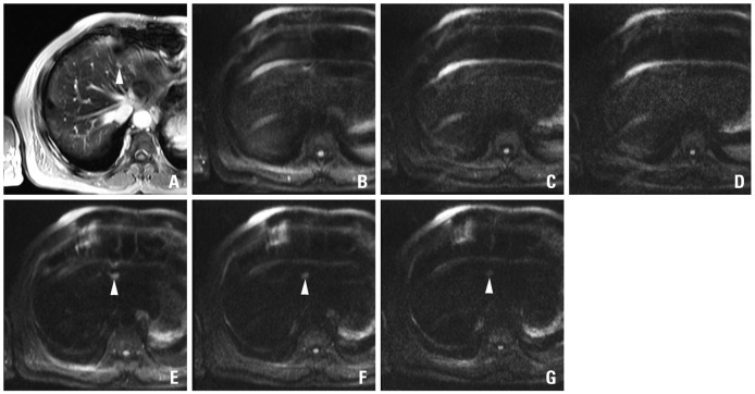
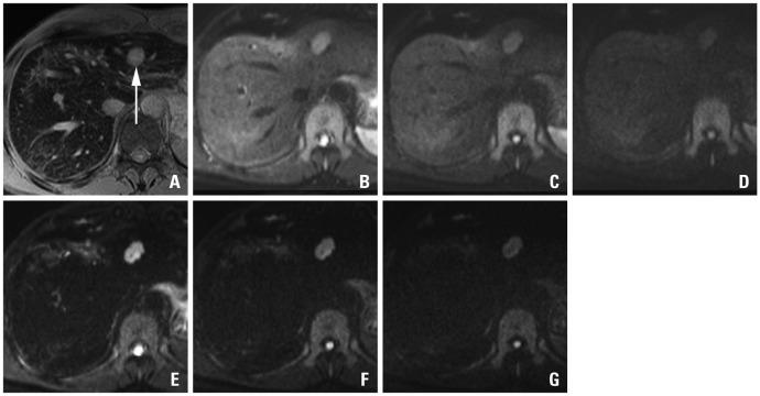
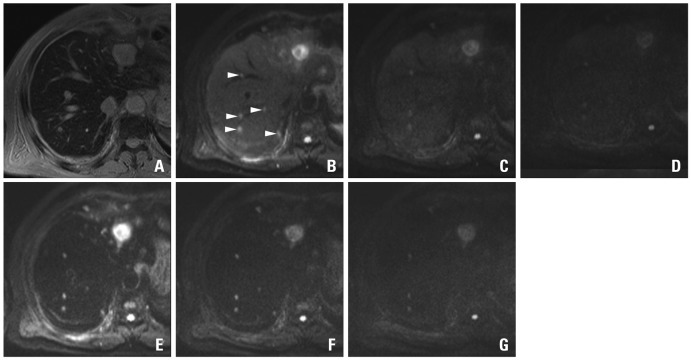
Eighty-six hepatic metastases (size range, 0.3-4.7 cm; mean, 1.5 cm) verified pathologically or by follow-up imaging studies in 22 consecutive patients (17 men and 5 women; 44-83 years; mean age, 60 years) during a 13-month period were enrolled. Hepatic MRI, including DWI (b-factors=50, 400, 800 s/mm²) with breath-holding technique of single-shot spin-echo echo-planar imaging (TR/TE=1000/69 ms, average=2) before and after SPIO administration, were retrospectively reviewed by two independent radiologists with a 5-point scale confidence score for each hepatic lesion on pre-contrast DWI (pre-DWI), SPIO-enhanced DWI (SPIO-DWI), and SPIO-enhanced T2*-weighted imaging (SPIO-T2*wI).

RESULTS

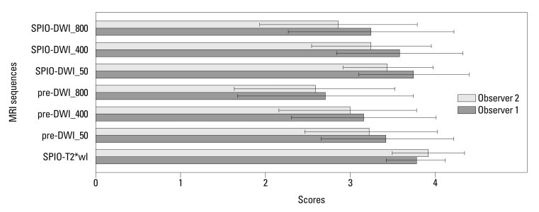
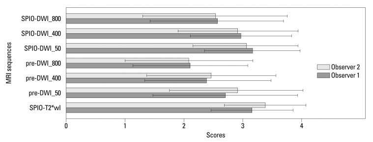
For all lesions, SPIO-T2wI showed significantly higher confidence score in the diagnosis of hepatic metastases than pre-contrast or SPIO-DWI regardless of the size of b-factors (p<0.05) with only one exception; using b-factor=50 s/mm², the score of SPIO-T2*wI was still higher than SPIO-DWI but there was no statistical significance given by observer 1 (p=0.730). For the subcentimeter lesions (n=37), SPIO-T2*wI showed the highest score, and using b-factor= 50 or 400 s/mm² SPIO-DWI showed similar confidence scores to SPIO-T2*wI by both observers (p>0.05). Pre-DWI using b-factor=50 sec/mm² was also comparable with SPIO-T2wI by observer 1 (p=0.060).

CONCLUSION

Pre-DWI has a limited value for the assessment of hepatic metastases, however, the repetition of DWI after SPIO injection using small b-factors could complement SPIO-T2*wI, especially for subcentimeter lesions.

摘要

目的

本研究的目的是验证超顺磁性氧化铁(SPIO)注射前后扩散加权 MRI(DWI)在评估肝转移中的作用。

材料与方法

连续 22 例(男 17 例,女 5 例;年龄 44-83 岁,平均 60 岁)患者的 86 个经病理或随访影像学检查证实的肝转移瘤(大小范围 0.3-4.7cm,平均 1.5cm)纳入研究。采用单次激发自旋回波平面成像呼吸门控技术进行肝脏 MRI 检查,包括 DWI(b 值=50、400、800s/mm²),并在 SPIO 给药前后进行(TR/TE=1000/69ms,平均 2 次)。两名独立的放射科医生对每个肝病变的平扫 DWI(pre-DWI)、SPIO 增强 DWI(SPIO-DWI)和 SPIO 增强 T2加权成像(SPIO-T2wI)的诊断信心进行五分制评分。

结果

所有病变中,SPIO-T2wI 在诊断肝转移瘤时的置信评分均显著高于平扫或 SPIO-DWI,与 b 值的大小无关(p<0.05),只有一个例外;使用 b 值=50s/mm² 时,SPIO-T2*wI 的评分仍高于 SPIO-DWI,但观察者 1 认为没有统计学意义(p=0.730)。对于亚厘米病变(n=37),SPIO-T2*wI 的评分最高,而观察者 1 认为使用 b 值=50 或 400s/mm² 的 SPIO-DWI 的置信评分与 SPIO-T2*wI 相似(p>0.05)。b 值=50sec/mm² 的平扫 DWI 与观察者 1 的 SPIO-T2wI 也相当(p=0.060)。

结论

平扫 DWI 对评估肝转移瘤的价值有限,然而,使用小 b 值重复 SPIO 注射后的 DWI 可以补充 SPIO-T2*wI,特别是对于亚厘米病变。

https://cdn.ncbi.nlm.nih.gov/pmc/blobs/695a/3381471/2c97bd62a730/ymj-53-825-g006.jpg
https://cdn.ncbi.nlm.nih.gov/pmc/blobs/695a/3381471/9028742b3dea/ymj-53-825-g001.jpg
https://cdn.ncbi.nlm.nih.gov/pmc/blobs/695a/3381471/1f469a39a81d/ymj-53-825-g002.jpg
https://cdn.ncbi.nlm.nih.gov/pmc/blobs/695a/3381471/8c9826ac06b3/ymj-53-825-g003.jpg
https://cdn.ncbi.nlm.nih.gov/pmc/blobs/695a/3381471/0b97fbbbecae/ymj-53-825-g004.jpg
https://cdn.ncbi.nlm.nih.gov/pmc/blobs/695a/3381471/16c742054178/ymj-53-825-g005.jpg
https://cdn.ncbi.nlm.nih.gov/pmc/blobs/695a/3381471/2c97bd62a730/ymj-53-825-g006.jpg

相似文献

1
Diffusion-weighted MR imaging before and after contrast enhancement with superparamagnetic iron oxide for assessment of hepatic metastasis.超顺磁性氧化铁对比增强前后弥散加权磁共振成像在肝转移评估中的应用。
Yonsei Med J. 2012 Jul 1;53(4):825-33. doi: 10.3349/ymj.2012.53.4.825.
2
Hepatic metastases: diffusion-weighted sensitivity-encoding versus SPIO-enhanced MR imaging.肝转移瘤:扩散加权敏感性编码与超顺磁性氧化铁增强磁共振成像
Radiology. 2006 Apr;239(1):122-30. doi: 10.1148/radiol.2383041384. Epub 2006 Feb 21.
3
Evaluation of super paramagnetic iron oxide-enhanced diffusion-weighted PROPELLER T2-fast spin echo magnetic resonance imaging: Preliminary experience.超顺磁性氧化铁增强扩散加权螺旋桨T2快速自旋回波磁共振成像的评估:初步经验。
J Comput Assist Tomogr. 2006 Mar-Apr;30(2):197-200. doi: 10.1097/00004728-200603000-00005.
4
Hypervascular hepatocellular carcinoma in the cirrhotic liver: diffusion-weighted imaging versus superparamagnetic iron oxide-enhanced MRI.肝硬化肝脏中的富血管性肝细胞癌:弥散加权成像与超顺磁性氧化铁增强 MRI 的比较。
Magn Reson Imaging. 2011 Nov;29(9):1235-43. doi: 10.1016/j.mri.2011.07.025. Epub 2011 Sep 9.
5
Superparamagnetic iron-oxide-enhanced diffusion-weighted magnetic resonance imaging for the diagnosis of intrapancreatic accessory spleen.超顺磁性氧化铁增强弥散加权磁共振成像在胰内副脾诊断中的应用。
Abdom Radiol (NY). 2019 Oct;44(10):3325-3335. doi: 10.1007/s00261-019-02189-8.
6
Detection of hepatocellular carcinoma (HCC) using super paramagnetic iron oxide (SPIO)-enhanced MRI: Added value of diffusion-weighted imaging (DWI).超顺磁性氧化铁(SPIO)增强 MRI 检测肝细胞癌(HCC):扩散加权成像(DWI)的附加价值。
J Magn Reson Imaging. 2010 Feb;31(2):373-82. doi: 10.1002/jmri.22059.
7
Hepatocellular lesions with increased iron uptake on superparamagnetic iron oxide-enhanced magnetic resonance imaging in cirrhosis or chronic hepatitis: comparison of four magnetic resonance sequences for lesion conspicuity.肝硬化或慢性肝炎患者中,超顺磁性氧化铁增强磁共振成像显示铁摄取增加的肝细胞病变:四种磁共振序列对病变显示清晰度的比较
Magn Reson Imaging. 2009 Jul;27(6):801-6. doi: 10.1016/j.mri.2008.11.006. Epub 2009 Jan 13.
8
Diffusion-weighted imaging versus superparamagnetic iron oxide (SPIO)-enhanced MRI: exclusive and combined values in the assessment of hepatic metastases.弥散加权成像与超顺磁性氧化铁(SPIO)增强 MRI 比较:在评估肝转移瘤中的单独和联合价值。
Magn Reson Imaging. 2012 May;30(4):554-61. doi: 10.1016/j.mri.2011.12.007. Epub 2012 Feb 6.
9
[Hepatic and hepatocarcinoma magnetic resonance: comparison of the results obtained with paramagnetic (gadolinium) and superparamagnetic (iron oxide particles) contrast media].[肝脏及肝癌的磁共振成像:顺磁性(钆)与超顺磁性(氧化铁颗粒)对比剂成像结果比较]
Radiol Med. 2000 Sep;100(3):160-7.
10
Colorectal hepatic metastases: detection with SPIO-enhanced breath-hold MR imaging--comparison of optimized sequences.结直肠癌肝转移:利用超顺磁性氧化铁增强屏气磁共振成像检测——优化序列的比较
Radiology. 2003 Sep;228(3):709-18. doi: 10.1148/radiol.2283020376. Epub 2003 Jul 29.

引用本文的文献

1
How PET/MR Can Add Value For Children With Cancer.PET/MR如何为癌症患儿增添价值。
Curr Radiol Rep. 2017 Mar;5(3). doi: 10.1007/s40134-017-0207-y. Epub 2017 Feb 21.
2
Nanosized contrast agents to noninvasively detect kidney inflammation by magnetic resonance imaging.纳米级对比剂通过磁共振成像非侵入性检测肾脏炎症。
Adv Chronic Kidney Dis. 2013 Nov;20(6):488-99. doi: 10.1053/j.ackd.2013.06.001.

本文引用的文献

1
Diffusion-weighted MR imaging of the liver.肝脏弥散加权磁共振成像。
Radiology. 2010 Jan;254(1):47-66. doi: 10.1148/radiol.09090021.
2
Diffusion-weighted magnetic resonance imaging for characterization of focal liver masses: impact of parallel imaging (SENSE) and b value.用于肝脏局灶性肿块特征分析的扩散加权磁共振成像:并行成像(敏感性编码技术)和b值的影响
J Comput Assist Tomogr. 2008 Nov-Dec;32(6):865-71. doi: 10.1097/RCT.0b013e3181591cf2.
3
Focal liver lesion detection and characterization: comparison of non-contrast enhanced and SPIO-enhanced diffusion-weighted single-shot spin echo echo planar and turbo spin echo T2-weighted imaging.
肝脏局灶性病变的检出与定性:非增强对比剂与超顺磁性氧化铁增强扩散加权单次激发自旋回波平面回波和涡轮自旋回波 T2 加权成像的比较。
Eur J Radiol. 2009 Dec;72(3):432-9. doi: 10.1016/j.ejrad.2008.09.005. Epub 2008 Oct 11.
4
Focal liver lesion detection and characterization with diffusion-weighted MR imaging: comparison with standard breath-hold T2-weighted imaging.磁共振扩散加权成像对肝脏局灶性病变的检测与特征分析:与标准屏气T2加权成像的比较
Radiology. 2008 Mar;246(3):812-22. doi: 10.1148/radiol.2463070432. Epub 2008 Jan 25.
5
Single breath-hold diffusion-weighted MRI of the liver with parallel imaging: initial experience.肝脏单次屏气并行成像扩散加权磁共振成像:初步经验
Clin Radiol. 2006 Nov;61(11):959-65. doi: 10.1016/j.crad.2006.06.014.
6
New MR techniques for the detection of liver metastases.用于检测肝转移瘤的新型磁共振技术。
Cancer Imaging. 2006 May 31;6(1):33-42. doi: 10.1102/1470-7330.2006.0007.
7
Evaluation of super paramagnetic iron oxide-enhanced diffusion-weighted PROPELLER T2-fast spin echo magnetic resonance imaging: Preliminary experience.超顺磁性氧化铁增强扩散加权螺旋桨T2快速自旋回波磁共振成像的评估:初步经验。
J Comput Assist Tomogr. 2006 Mar-Apr;30(2):197-200. doi: 10.1097/00004728-200603000-00005.
8
Hepatic metastases: diffusion-weighted sensitivity-encoding versus SPIO-enhanced MR imaging.肝转移瘤:扩散加权敏感性编码与超顺磁性氧化铁增强磁共振成像
Radiology. 2006 Apr;239(1):122-30. doi: 10.1148/radiol.2383041384. Epub 2006 Feb 21.
9
Diffusion-weighted images of the liver: comparison of tumor detection before and after contrast enhancement with superparamagnetic iron oxide.肝脏的扩散加权成像:超顺磁性氧化铁增强前后肿瘤检测的比较。
J Magn Reson Imaging. 2005 Jun;21(6):836-40. doi: 10.1002/jmri.20346.
10
Can a single-shot black-blood T2-weighted spin-echo echo-planar imaging sequence with sensitivity encoding replace the respiratory-triggered turbo spin-echo sequence for the liver? An optimization and feasibility study.具有敏感性编码的单次激发黑血T2加权自旋回波平面成像序列能否替代肝脏的呼吸触发涡轮自旋回波序列?一项优化与可行性研究。
J Magn Reson Imaging. 2005 Mar;21(3):219-29. doi: 10.1002/jmri.20269.