Hamdoon Zaid, Jerjes Waseem, Al-Delayme Raed, McKenzie Gordon, Jay Amrita, Hopper Colin
Department of Oral & Maxillofacial Surgery, Dental School, Mosul, Iraq.
Head Neck Oncol. 2012 Jun 6;4:29. doi: 10.1186/1758-3284-4-29.
Optical coherence tomography (OCT) is a non-invasive optical technology using near-infrared light to produce cross-sectional tissue images with lateral resolution.
The overall aims of this study was to generate a bank of normative and pathological OCT data of the oral tissues to allow identification of cellular structures of normal and pathological processes with the aim to create a diagnostic algorithm which can be used in the early detection of oral disorders.
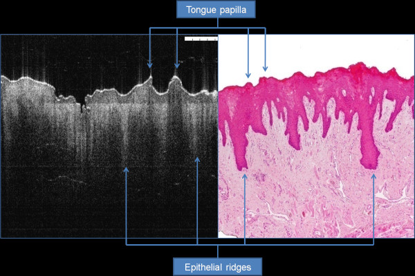
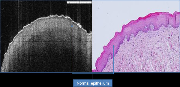
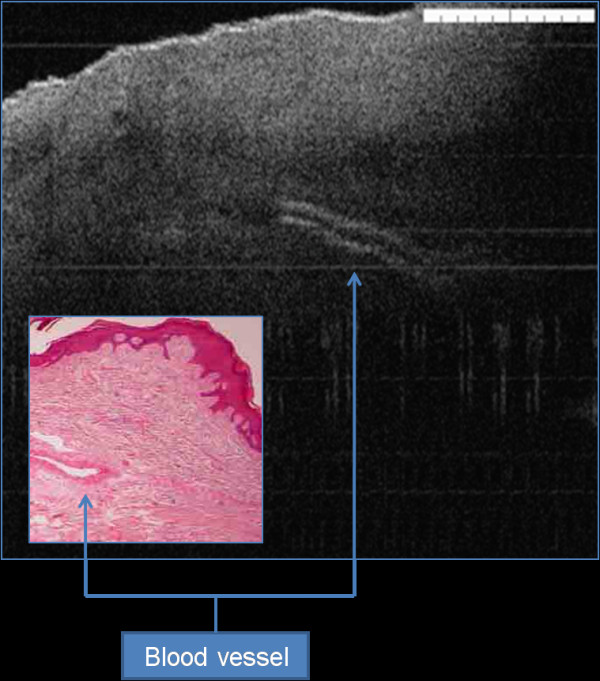
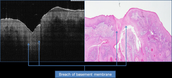
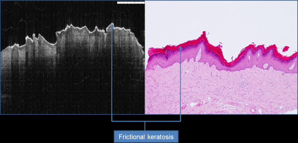
Seventy-three patients with 78 suspicious oral lesions were referred for further management to the UCLH Head and Neck Centre, London. The entire cohort had their lesions surgically biopsied (incisional or excisional). The immediate ex vivo phase involved scanning the specimens using optical coherence tomography. The specimens were then processed by a histopathologist. Five tissue structures were evaluated as part of this study, including: keratin cell layer, epithelial layer, basement membrane, lamina propria and other microanatomical structures. Two independent assessors (clinician and pathologist trained to use OCT) assessed the OCT images and were asked to comment on the cellular structures and changes involving the five tissue structures in non-blind fashion.
Correct identification of the keratin cell layer and its structural changes was achieved in 87% of the cohort; for the epithelial layer it reached 93.5%, and 94% for the basement membrane. Microanatomical structures identification was 64% for blood vessels, 58% for salivary gland ducts and 89% for rete pegs. The agreement was "good" between the clinician and the pathologist. OCT was able to differential normal from pathological tissue and pathological tissue of different entities in this immediate ex vivo study. Unfortunately, OCT provided inadequate cellular and subcellular information to enable the grading of oral premalignant disorders.
This study enabled the creation of OCT bank of normal and pathological oral tissues. The pathological changes identified using OCT enabled differentiation between normal and pathological tissues, and identification of different tissue pathologies. Further studies are required to assess the accuracy of OCT in identification of various pathological processes involving the oral tissues.
光学相干断层扫描(OCT)是一种非侵入性光学技术,利用近红外光生成具有横向分辨率的组织横截面图像。
本研究的总体目标是建立一组口腔组织的正常和病理OCT数据,以便识别正常和病理过程中的细胞结构,旨在创建一种可用于口腔疾病早期检测的诊断算法。
73例患有78个可疑口腔病变的患者被转诊至伦敦大学学院医院头颈中心进行进一步治疗。整个队列的病变均接受手术活检(切取或切除)。离体即刻阶段包括使用光学相干断层扫描对标本进行扫描。然后由组织病理学家对标本进行处理。作为本研究的一部分,评估了五种组织结构,包括:角质形成细胞层、上皮层、基底膜、固有层和其他微观解剖结构。两名独立评估者(经过使用OCT培训的临床医生和病理学家)对OCT图像进行评估,并被要求以非盲法对涉及五种组织结构的细胞结构和变化进行评论。
在87%的队列中正确识别了角质形成细胞层及其结构变化;上皮层的识别率达到93.5%,基底膜的识别率为94%。微观解剖结构的识别率为:血管64%,唾液腺导管58%,钉突89%。临床医生和病理学家之间的一致性“良好”。在本离体即刻研究中,OCT能够区分正常组织与病理组织以及不同实体的病理组织。不幸的是,OCT提供的细胞和亚细胞信息不足,无法对口腔癌前病变进行分级。
本研究建立了正常和病理口腔组织的OCT数据库。使用OCT识别出的病理变化能够区分正常组织与病理组织,并识别不同的组织病理学。需要进一步研究以评估OCT在识别涉及口腔组织的各种病理过程中的准确性。