Division of Infectious Diseases, Department of Medicine, Hospital Sao Paulo, Escola Paulista de Medicina, Universidade Federal de Sao Paulo, Sao Paulo, Brazil.
PLoS One. 2012;7(5):e37393. doi: 10.1371/journal.pone.0037393. Epub 2012 May 31.
A shift from Th1 to Th2 as well as an increase in Treg CD4+T cell subsets has been reported in septic patients (SP). Furthermore, these patients display modulation of monocyte function, with reduced production of pro-inflammatory cytokines upon LPS stimulus, which resembles the phenotype of alternatively activated macrophages. In this study, we evaluated the percentages of T cells differentiated into Th1, Th17 and Treg subsets, as well as the percentage of monocytes expressing markers of alternatively activated monocytes/macrophages (AAM) in SP.
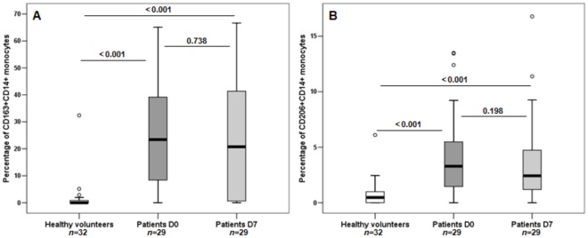
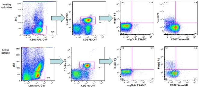
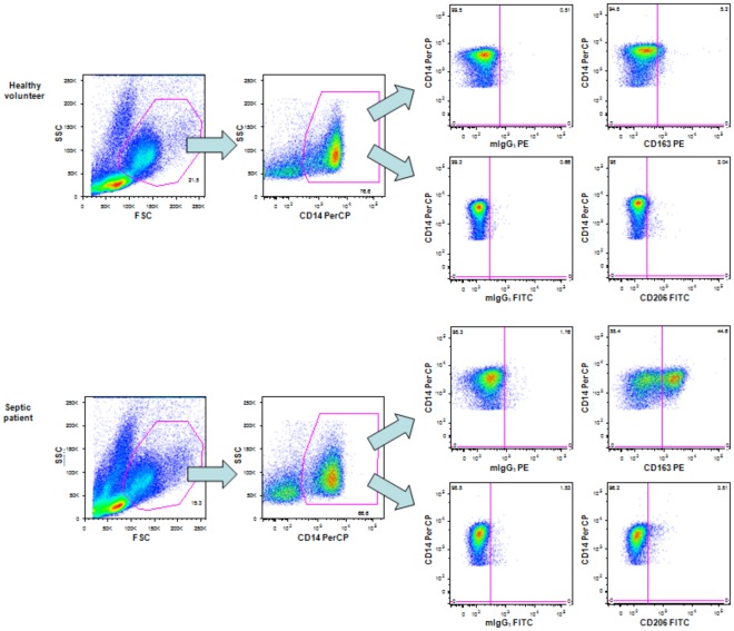
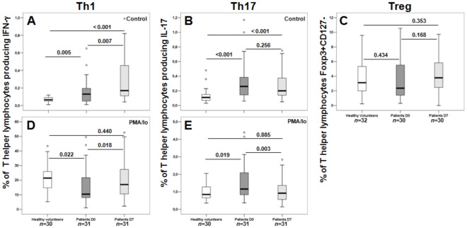
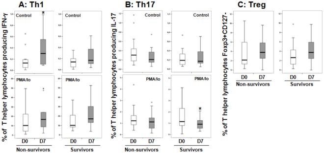
METHODOLOGY/PRINCIPAL FINDINGS: Peripheral blood mononuclear cells (PBMC) were obtained from 32 healthy volunteers (HV) and from SP at admission (D0, n = 67) and after 7 days of therapy (D7, n = 33). Th1 and Th17 (CD3+CD8-) lymphocytes were identified by the intracellular detection of IFN-γ and IL-17, respectively, spontaneously and after PMA/Io stimulation, and Treg cells were identified by Foxp3+CD127- expression. Monocytes were evaluated for CD206 and CD163 expression. Absolute numbers of CD4+T lymphocytes were measured in whole blood samples by flow cytometry. The Mann-Whitney or Wilcoxon test was applied, as appropriate. The percentage of Th1 cells was lower in SP than in HV at admission after PMA/Io stimulation, whereas the percentage of Th17 cells was higher. In patients' follow-up samples, a higher percentage of Th1 cells and a lower percentage of Th17 cells were observed on D7 compared with the D0 samples. Treg cells remained unchanged. Septic patients showed a markedly increased proportion of monocytes expressing CD163 and CD206.
CONCLUSIONS/SIGNIFICANCE: Upon in vitro stimulus, the percentage of T helper lymphocytes producing IL-17 was higher in SP than in HV at admission, and the percentage producing IFN-γ was lower, a pattern that was reversed during follow-up. The increased expression of CD163 and CD206 indicates that monocytes may acquire the AAM phenotype during sepsis.
据报道,脓毒症患者(SP)体内 Th1 向 Th2 的转变以及 Treg CD4+T 细胞亚群的增加。此外,这些患者表现出单核细胞功能的调节,即在 LPS 刺激下产生促炎细胞因子的能力降低,这类似于替代激活的巨噬细胞表型。在这项研究中,我们评估了 Th1、Th17 和 Treg 细胞亚群分化的 T 细胞的百分比,以及在 SP 中表达替代激活的单核细胞/巨噬细胞(AAM)标志物的单核细胞的百分比。
方法/主要发现:从 32 名健康志愿者(HV)和 SP 入院时(D0,n=67)和治疗 7 天后(D7,n=33)获得外周血单核细胞(PBMC)。通过 IFN-γ 和 IL-17 的细胞内检测分别鉴定 Th1 和 Th17(CD3+CD8-)淋巴细胞,自发和 PMA/Io 刺激后,并通过 Foxp3+CD127-表达鉴定 Treg 细胞。评估单核细胞的 CD206 和 CD163 表达。通过流式细胞术在全血样本中测量 CD4+T 淋巴细胞的绝对数量。应用 Mann-Whitney 或 Wilcoxon 检验,视情况而定。在 PMA/Io 刺激后,SP 患者入院时的 Th1 细胞百分比低于 HV,而 Th17 细胞百分比较高。在患者的随访样本中,与 D0 样本相比,D7 时观察到 Th1 细胞的百分比较高,Th17 细胞的百分比较低。Treg 细胞保持不变。脓毒症患者表现出表达 CD163 和 CD206 的单核细胞比例明显增加。
结论/意义:在体外刺激下,SP 患者入院时产生 IL-17 的辅助性 T 淋巴细胞的百分比高于 HV,而产生 IFN-γ 的百分比较低,这种模式在随访期间发生逆转。CD163 和 CD206 的表达增加表明单核细胞在脓毒症期间可能获得 AAM 表型。