Pennsylvania Muscle Institute, School of Medicine, University of Pennsylvania, Philadelphia, Pennsylvania, United States of America.
PLoS One. 2012;7(5):e38344. doi: 10.1371/journal.pone.0038344. Epub 2012 May 31.
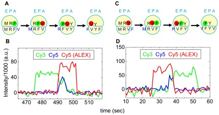
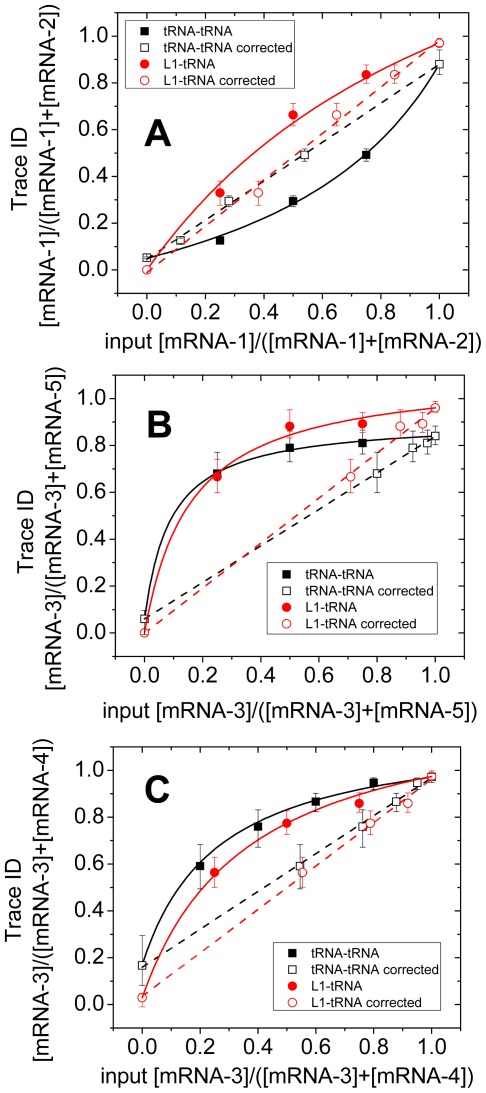
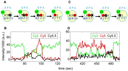
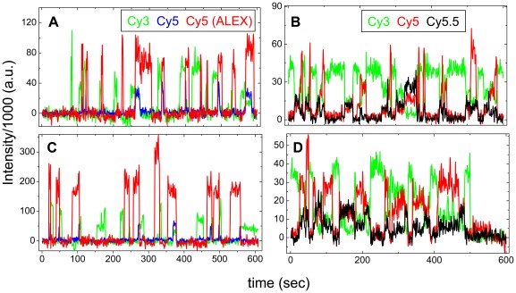
We present proof-of-concept in vitro results demonstrating the feasibility of using single molecule fluorescence resonance energy transfer (smFRET) measurements to distinguish, in real time, between individual ribosomes programmed with several different, short mRNAs. For these measurements we use either the FRET signal generated between two tRNAs labeled with different fluorophores bound simultaneously in adjacent sites to the ribosome (tRNA-tRNA FRET) or the FRET signal generated between a labeled tRNA bound to the ribosome and a fluorescent derivative of ribosomal protein L1 (L1-tRNA FRET). With either technique, criteria were developed to identify the mRNAs, taking into account the relative activity of the mRNAs. These criteria enabled identification of the mRNA being translated by a given ribosome to within 95% confidence intervals based on the number of identified FRET traces. To upgrade the approach for natural mRNAs or more complex mixtures, the stoichiometry of labeling should be enhanced and photobleaching reduced. The potential for porting these methods into living cells is discussed.
我们提出了体外概念验证结果,证明使用单分子荧光共振能量转移(smFRET)测量实时区分核糖体上不同短 mRNA 的可行性。对于这些测量,我们使用两种 FRET 信号:一种是由两个标记有不同荧光团的 tRNA 在核糖体的相邻位置同时结合产生的(tRNA-tRNA FRET),另一种是由核糖体上结合的标记 tRNA 和核糖体蛋白 L1 的荧光衍生物产生的(L1-tRNA FRET)。对于这两种技术,我们制定了考虑到 mRNA 相对活性的标准来识别 mRNA。这些标准使我们能够根据识别的 FRET 痕迹的数量,在 95%置信区间内识别给定核糖体正在翻译的 mRNA。为了将这些方法升级到天然 mRNA 或更复杂的混合物,应增强标记的化学计量并减少光漂白。讨论了将这些方法移植到活细胞的可能性。