Reproductive Centre, Mio Fertility Clinic, 2-1-1 Kuzumo-Minami, Yonago, Japan.
J Assist Reprod Genet. 2012 Sep;29(9):951-6. doi: 10.1007/s10815-012-9815-x. Epub 2012 Jun 14.
To analyze the fertilization process related to polyspermy block in human oocytes using an in vitro culturing system for time-lapse cinematography.
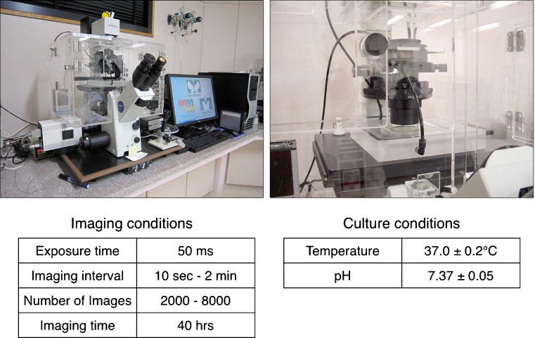
We had 122 oocytes donated for this study from couples that provided informed consent. We recorded human oocytes at 2,000 to 2,800 frames every 10 s during the fertilization process and thereafter every 2 min using a new in vitro culture system originally developed by the authors for time-lapse cinematography. We displayed 30 frames per second for analysis of the polyspermy block during fertilization.
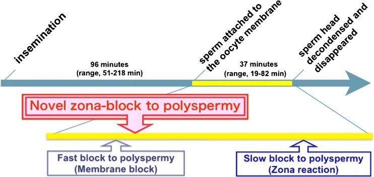
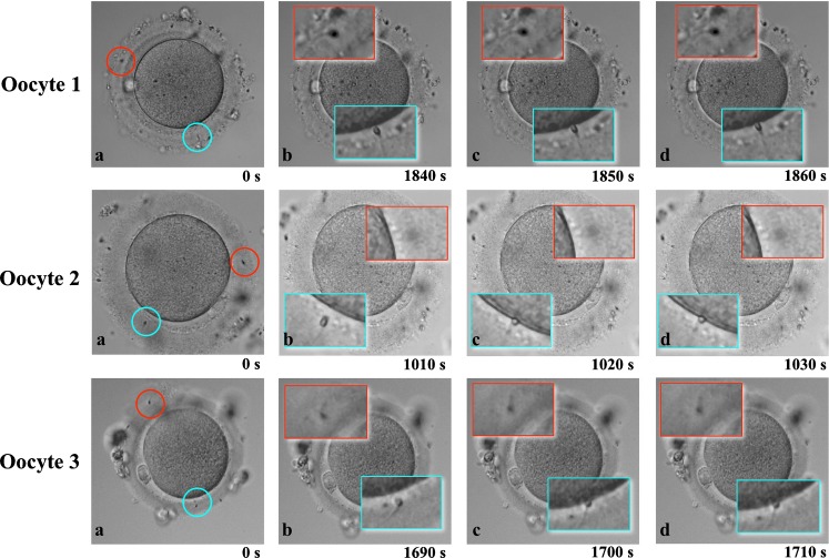
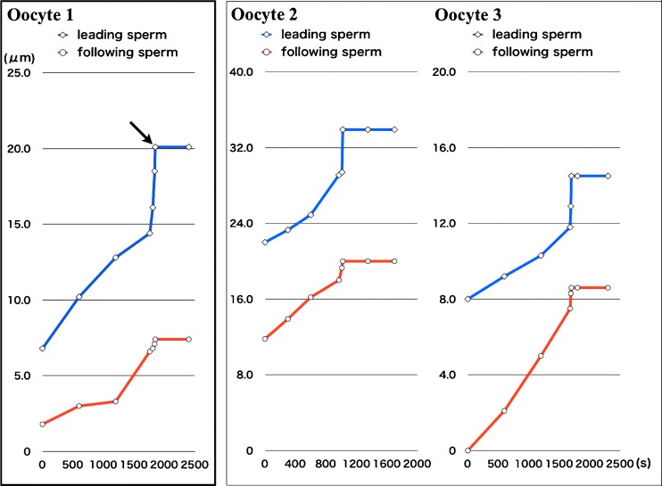
Three oocytes showed the leading and following sperm within the zona pellucida in the same microscopic field. The dynamic images obtained during the fertilization process using this new system revealed that once a leading sperm penetrated the zona pellucida and attached to the oocyte membrane, a following sperm was arrested from further penetration into the zona pellucida within 10 s.
The present results strongly suggest the existence of a novel mechanism of polyspermy block that takes place at the zona pellucida immediately after fertilization. These findings are clearly different from previous mechanisms describing polyspermy block as the oocyte membrane block to sperm penetration and the zona reaction. The finding presented herein thus represents a novel discovery about the highly complicated polyspermy block mechanism occurring in human oocytes.
利用体外延时摄像培养系统分析人卵多精受精阻滞过程。
本研究共收集了 122 枚经知情同意后自愿捐献的卵母细胞。我们使用作者新开发的体外延时摄像培养系统,以每 10 秒 2000-2800 帧的速度记录受精过程中的人卵,并在此后每 2 分钟记录一次。我们以每秒 30 帧的速度分析受精过程中的多精受精阻滞。
在同一显微镜视野下,有 3 枚卵母细胞显示透明带内有前导精子和后续精子。使用新系统获得的受精过程动态图像显示,一旦前导精子穿透透明带并附着在卵母细胞膜上,后续精子在 10 秒内被阻止进一步穿透透明带。
本研究结果强烈提示在受精后立即发生的透明带处存在一种新的多精受精阻滞机制。这些发现与之前描述的多精受精阻滞机制明显不同,后者将卵母细胞膜阻滞精子穿透和透明带反应作为多精受精阻滞的机制。因此,本文的发现代表了人类卵母细胞中高度复杂的多精受精阻滞机制的一个新发现。