• 文献检索
  • 文档翻译
  • 深度研究
  • 学术资讯
  • Suppr Zotero 插件Zotero 插件
  • 邀请有礼
  • 套餐&价格
  • 历史记录
应用&插件
Suppr Zotero 插件Zotero 插件浏览器插件Mac 客户端Windows 客户端微信小程序
定价
高级版会员购买积分包购买API积分包
服务
文献检索文档翻译深度研究API 文档MCP 服务
关于我们
关于 Suppr公司介绍联系我们用户协议隐私条款
关注我们

Suppr 超能文献

核心技术专利:CN118964589B侵权必究
粤ICP备2023148730 号-1Suppr @ 2026

文献检索

告别复杂PubMed语法,用中文像聊天一样搜索,搜遍4000万医学文献。AI智能推荐,让科研检索更轻松。

立即免费搜索

文件翻译

保留排版,准确专业,支持PDF/Word/PPT等文件格式,支持 12+语言互译。

免费翻译文档

深度研究

AI帮你快速写综述,25分钟生成高质量综述,智能提取关键信息,辅助科研写作。

立即免费体验

肺部磁共振成像(二/三)。为什么……何时……如何?

MRI of the lung (2/3). Why … when … how?

机构信息

University Hospital Schleswig-Holstein, Campus Kiel, Department of Diagnostic Radiology, Arnold-Heller-Straße 3, Haus 23, 24105, Kiel, Germany,

出版信息

Insights Imaging. 2012 Aug;3(4):355-71. doi: 10.1007/s13244-011-0146-8. Epub 2012 Feb 13.

DOI:10.1007/s13244-011-0146-8
PMID:22695944
原文链接:https://pmc.ncbi.nlm.nih.gov/articles/PMC3481084/
Abstract

BACKGROUND

Among the modalities for lung imaging, proton magnetic resonance imaging (MRI) has been the latest to be introduced into clinical practice. Its value to replace X-ray and computed tomography (CT) when radiation exposure or iodinated contrast material is contra-indicated is well acknowledged: i.e. for paediatric patients and pregnant women or for scientific use. One of the reasons why MRI of the lung is still rarely used, except in a few centres, is the lack of consistent protocols customised to clinical needs.

METHODS

This article makes non-vendor-specific protocol suggestions for general use with state-of-the-art MRI scanners, based on the available literature and a consensus discussion within a panel of experts experienced in lung MRI.

RESULTS

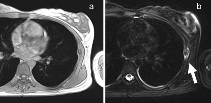
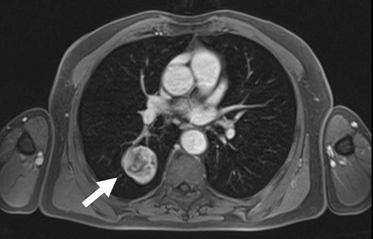
Various sequences have been successfully tested within scientific or clinical environments. MRI of the lung with appropriate combinations of these sequences comprises morphological and functional imaging aspects in a single examination. It serves in difficult clinical problems encountered in daily routine, such as assessment of the mediastinum and chest wall, and even might challenge molecular imaging techniques in the near future.

CONCLUSION

This article helps new users to implement appropriate protocols on their own MRI platforms. Main Messages • MRI of the lung can be readily performed on state-of-the-art 1.5-T MRI scanners. • Protocol suggestions based on the available literature facilitate its use for routine • MRI offers solutions for complicated thoracic masses with atelectasis and chest wall invasion. • MRI is an option for paediatrics and science when CT is contra-indicated.

摘要

背景

在肺部成像方式中,质子磁共振成像(MRI)是最新引入临床实践的一种。当存在辐射暴露或碘造影剂禁忌时,它可以替代 X 射线和计算机断层扫描(CT),这一点已得到广泛认可:例如,用于儿科患者、孕妇或科研用途。除了少数中心外,肺部 MRI 应用仍然较少的原因之一是缺乏针对临床需求定制的一致协议。

方法

本文基于现有文献和一组在肺部 MRI 方面经验丰富的专家的共识讨论,针对最先进的 MRI 扫描仪提出了非特定于供应商的通用协议建议。

结果

在科学或临床环境中已经成功测试了各种序列。通过适当组合这些序列,肺部 MRI 可以在单次检查中同时进行形态学和功能成像。它可用于解决日常工作中遇到的困难临床问题,例如评估纵隔和胸壁,甚至在不久的将来可能会挑战分子成像技术。

结论

本文有助于新用户在自己的 MRI 平台上实施适当的协议。主要信息

  1. 肺部 MRI 可以在最先进的 1.5-T MRI 扫描仪上轻松进行。

  2. 基于现有文献的协议建议有助于常规应用。

  3. MRI 为伴有肺不张和胸壁侵犯的复杂胸部肿块提供了解决方案。

  4. 在 CT 禁忌的情况下,MRI 是儿科和科研的一种选择。

https://cdn.ncbi.nlm.nih.gov/pmc/blobs/969e/3481084/19136bfff432/13244_2011_146_Fig10_HTML.jpg
https://cdn.ncbi.nlm.nih.gov/pmc/blobs/969e/3481084/74a1b506aed2/13244_2011_146_Fig1_HTML.jpg
https://cdn.ncbi.nlm.nih.gov/pmc/blobs/969e/3481084/635df5bac8b8/13244_2011_146_Fig2_HTML.jpg
https://cdn.ncbi.nlm.nih.gov/pmc/blobs/969e/3481084/9e8eef7514dd/13244_2011_146_Fig3_HTML.jpg
https://cdn.ncbi.nlm.nih.gov/pmc/blobs/969e/3481084/e5205c368f85/13244_2011_146_Fig4_HTML.jpg
https://cdn.ncbi.nlm.nih.gov/pmc/blobs/969e/3481084/9bbd4f5d7de4/13244_2011_146_Fig5_HTML.jpg
https://cdn.ncbi.nlm.nih.gov/pmc/blobs/969e/3481084/bad245e5c0f2/13244_2011_146_Fig6_HTML.jpg
https://cdn.ncbi.nlm.nih.gov/pmc/blobs/969e/3481084/806c691029ab/13244_2011_146_Fig7_HTML.jpg
https://cdn.ncbi.nlm.nih.gov/pmc/blobs/969e/3481084/24b9094d504f/13244_2011_146_Fig8_HTML.jpg
https://cdn.ncbi.nlm.nih.gov/pmc/blobs/969e/3481084/06fbbc60d1b4/13244_2011_146_Fig9_HTML.jpg
https://cdn.ncbi.nlm.nih.gov/pmc/blobs/969e/3481084/19136bfff432/13244_2011_146_Fig10_HTML.jpg

相似文献

1
MRI of the lung (2/3). Why … when … how?肺部磁共振成像(二/三)。为什么……何时……如何?
Insights Imaging. 2012 Aug;3(4):355-71. doi: 10.1007/s13244-011-0146-8. Epub 2012 Feb 13.
2
MRI of the lung (3/3)-current applications and future perspectives.肺部 MRI(3/3)——当前的应用与未来展望。
Insights Imaging. 2012 Aug;3(4):373-86. doi: 10.1007/s13244-011-0142-z. Epub 2012 Jan 15.
3
Variabilities of Magnetic Resonance Imaging-, Computed Tomography-, and Positron Emission Tomography-Computed Tomography-Based Tumor and Lymph Node Delineations for Lung Cancer Radiation Therapy Planning.基于磁共振成像、计算机断层扫描以及正电子发射断层扫描-计算机断层扫描的肺癌放射治疗计划中肿瘤及淋巴结勾画的变异性
Int J Radiat Oncol Biol Phys. 2017 Sep 1;99(1):80-89. doi: 10.1016/j.ijrobp.2017.05.002. Epub 2017 May 6.
4
[MRI of interstitial lung diseases: what is possible?].[间质性肺疾病的磁共振成像:能实现什么?]
Radiologe. 2014 Dec;54(12):1204-12. doi: 10.1007/s00117-014-2738-z.
5
Ten years of chest MRI for patients with cystic fibrosis : Translation from the bench to clinical routine.针对囊性纤维化患者的十年胸部磁共振成像:从实验台到临床常规应用的转化
Radiologe. 2019 Dec;59(Suppl 1):10-20. doi: 10.1007/s00117-019-0553-2.
6
Trial design and reporting standards for intra-arterial cerebral thrombolysis for acute ischemic stroke.急性缺血性脑卒中动脉内脑溶栓的试验设计与报告标准。
Stroke. 2003 Aug;34(8):e109-37. doi: 10.1161/01.STR.0000082721.62796.09. Epub 2003 Jul 17.
7
Assessment of morphological MRI for pulmonary changes in cystic fibrosis (CF) patients: comparison to thin-section CT and chest x-ray.囊性纤维化(CF)患者肺部改变的形态学磁共振成像评估:与薄层CT和胸部X线的比较
Invest Radiol. 2007 Oct;42(10):715-25. doi: 10.1097/RLI.0b013e318074fd81.
8
Pediatric Chest MRI: A Review.儿科胸部 MRI:综述。
Indian J Pediatr. 2019 Sep;86(9):842-853. doi: 10.1007/s12098-018-02852-w. Epub 2019 Feb 5.
9
MRI of the lung: state of the art.肺部磁共振成像:最新技术。
Diagn Interv Radiol. 2012 Jul-Aug;18(4):344-53. doi: 10.4261/1305-3825.DIR.5365-11.0. Epub 2012 Mar 20.
10
New imaging techniques in the treatment guidelines for lung cancer.肺癌治疗指南中的新成像技术。
Eur Respir J Suppl. 2002 Feb;35:71s-83s. doi: 10.1183/09031936.02.00277902.

引用本文的文献

1
Non-contrast enhanced functional lung MRI in children: systematic review.儿童非对比增强功能性肺部磁共振成像:系统评价
Front Pediatr. 2025 Jul 22;13:1568172. doi: 10.3389/fped.2025.1568172. eCollection 2025.
2
[Magnetic resonance imaging of the pediatric chest].[小儿胸部的磁共振成像]
Radiologie (Heidelb). 2025 Jul 9. doi: 10.1007/s00117-025-01486-2.
3
18F-FDG PET and combined diffusion weighted MRI features in pleural infection: A case report.18F-FDG PET与联合扩散加权磁共振成像特征在胸膜感染中的应用:一例报告

本文引用的文献

1
Diffusion-weighted MR-imaging for the detection of pulmonary nodules at 1.5 Tesla: intraindividual comparison with multidetector computed tomography.1.5特斯拉磁共振成像扩散加权成像用于检测肺结节:与多排螺旋计算机断层扫描的个体内比较
J Med Imaging Radiat Oncol. 2011 Jun;55(3):266-74. doi: 10.1111/j.1754-9485.2011.02263.x.
2
Lung MRI as a possible alternative to CT scan for patients with primary immune deficiencies and increased radiosensitivity.肺部 MRI 可能成为原发性免疫缺陷和辐射敏感性增加患者的 CT 扫描替代方法。
Chest. 2011 Dec;140(6):1581-1589. doi: 10.1378/chest.10-3147. Epub 2011 May 26.
3
Pulmonary functional imaging: qualitative comparison of Fourier decomposition MR imaging with SPECT/CT in porcine lung.
Radiol Case Rep. 2025 Jun 21;20(9):4492-4496. doi: 10.1016/j.radcr.2025.05.083. eCollection 2025 Sep.
4
The value of multisequence MR in the morphological evaluation of peripheral lung cancer.多序列磁共振成像在外周型肺癌形态学评估中的价值
BMC Med Imaging. 2025 May 21;25(1):178. doi: 10.1186/s12880-025-01726-w.
5
Non-contrast enhanced functional lung MRI in children: report on 900 own measurements using matrix-pencil decomposition (MP-) MRI.儿童非对比增强功能性肺部磁共振成像:基于900例自身测量的矩阵铅笔分解(MP-)MRI报告
Front Pediatr. 2025 Mar 13;13:1519148. doi: 10.3389/fped.2025.1519148. eCollection 2025.
6
Post-correctional improvement of T2-weighted fast spin echo magnetic resonance imaging pulse sequence for detecting high intensity focused ultrasound thermal lesions.用于检测高强度聚焦超声热损伤的T2加权快速自旋回波磁共振成像脉冲序列的校正后改进
Heliyon. 2024 Nov 16;10(22):e40496. doi: 10.1016/j.heliyon.2024.e40496. eCollection 2024 Nov 30.
7
Using machine learning for early detection of chronic obstructive pulmonary disease: a narrative review.利用机器学习进行慢性阻塞性肺疾病的早期检测:叙述性综述。
Respir Res. 2024 Sep 9;25(1):336. doi: 10.1186/s12931-024-02960-6.
8
Deep learning applications for quantitative and qualitative PET in PET/MR: technical and clinical unmet needs.深度学习在 PET/MR 中的定量和定性 PET 中的应用:技术和临床未满足的需求。
MAGMA. 2024 Aug;37(4):749-763. doi: 10.1007/s10334-024-01199-y. Epub 2024 Aug 21.
9
Characterizing Lung Parenchymal Aeration via Standardized Signal Intensity from Free-breathing 4D Dynamic MRI in Phantoms, Healthy Children, and Pediatric Patients with Thoracic Insufficiency Syndrome.通过自由呼吸 4D 动态 MRI 对肺实质充气进行特征描述:在体模、健康儿童和患有胸壁顺应性降低综合征的儿科患者中的研究。
Radiol Cardiothorac Imaging. 2024 Aug;6(4):e230262. doi: 10.1148/ryct.230262.
10
Imaging the Lung in ARDS: A Primer.急性呼吸窘迫综合征肺部影像学:基础篇。
Respir Care. 2024 Jul 24;69(8):1011-1024. doi: 10.4187/respcare.12061.
肺部功能成像:傅里叶分解 MRI 与 SPECT/CT 在猪肺中的定性比较。
Radiology. 2011 Aug;260(2):551-9. doi: 10.1148/radiol.11102313. Epub 2011 May 17.
4
Morphologic and functional scoring of cystic fibrosis lung disease using MRI.采用 MRI 对囊性纤维化肺病进行形态学和功能学评分。
Eur J Radiol. 2012 Jun;81(6):1321-9. doi: 10.1016/j.ejrad.2011.02.045. Epub 2011 Mar 23.
5
Role of diffusion-weighted imaging in the differential diagnosis of benign and malignant lesions of the chest-mediastinum.弥散加权成像在鉴别胸部-纵膈良恶性病变中的作用。
Radiol Med. 2011 Aug;116(5):720-33. doi: 10.1007/s11547-011-0629-1. Epub 2011 Feb 1.
6
[Whole-body MRI in children and juveniles].[儿童和青少年的全身磁共振成像]
Rofo. 2011 Jan;183(1):24-36. doi: 10.1055/s-0029-1245883. Epub 2010 Dec 15.
7
Maximum intensity projection (MIP) imaging using slice-stacking MRI.采用层块叠加 MRI 的最大密度投影(MIP)成像。
Med Phys. 2010 Nov;37(11):5914-20. doi: 10.1118/1.3503850.
8
Diffusion-weighted MR imaging in comparison to integrated [¹⁸F]-FDG PET/CT for N-staging in patients with lung cancer.扩散加权磁共振成像与整合 [¹⁸F]-FDG PET/CT 在肺癌患者 N 分期中的比较。
Eur J Radiol. 2012 Jan;81(1):178-82. doi: 10.1016/j.ejrad.2010.09.001. Epub 2010 Oct 6.
9
Diffusion and perfusion MRI of the lung and mediastinum.肺部和纵隔的弥散和灌注 MRI。
Eur J Radiol. 2010 Dec;76(3):329-36. doi: 10.1016/j.ejrad.2010.05.005.
10
In vivo MRI of altered proton signal intensity and T2 relaxation in a bleomycin model of pulmonary inflammation and fibrosis.在博来霉素诱导的肺炎症和纤维化模型中,利用活体 MRI 观察质子信号强度和 T2 弛豫时间的改变。
J Magn Reson Imaging. 2010 May;31(5):1091-9. doi: 10.1002/jmri.22166.