• 文献检索
  • 文档翻译
  • 深度研究
  • 学术资讯
  • Suppr Zotero 插件Zotero 插件
  • 邀请有礼
  • 套餐&价格
  • 历史记录
应用&插件
Suppr Zotero 插件Zotero 插件浏览器插件Mac 客户端Windows 客户端微信小程序
定价
高级版会员购买积分包购买API积分包
服务
文献检索文档翻译深度研究API 文档MCP 服务
关于我们
关于 Suppr公司介绍联系我们用户协议隐私条款
关注我们

Suppr 超能文献

核心技术专利:CN118964589B侵权必究
粤ICP备2023148730 号-1Suppr @ 2026

文献检索

告别复杂PubMed语法,用中文像聊天一样搜索,搜遍4000万医学文献。AI智能推荐,让科研检索更轻松。

立即免费搜索

文件翻译

保留排版,准确专业,支持PDF/Word/PPT等文件格式,支持 12+语言互译。

免费翻译文档

深度研究

AI帮你快速写综述,25分钟生成高质量综述,智能提取关键信息,辅助科研写作。

立即免费体验

视网膜损伤模式有助于苏萨克综合征和 MS 的鉴别诊断。

Patterns of retinal damage facilitate differential diagnosis between Susac syndrome and MS.

机构信息

NeuroCure Clinical Research Center, Charité-Universitätsmedizin Berlin, Berlin, Germany.

出版信息

PLoS One. 2012;7(6):e38741. doi: 10.1371/journal.pone.0038741. Epub 2012 Jun 11.

DOI:10.1371/journal.pone.0038741
PMID:22701702
原文链接:https://pmc.ncbi.nlm.nih.gov/articles/PMC3372471/
Abstract

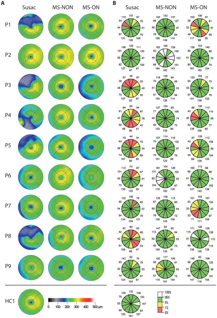
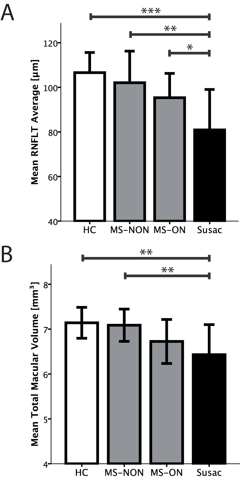
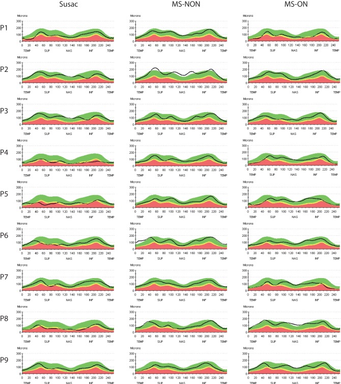
Susac syndrome, a rare but probably underdiagnosed combination of encephalopathy, hearing loss, and visual deficits due to branch retinal artery occlusion of unknown aetiology has to be considered as differential diagnosis in various conditions. Particularly, differentiation from multiple sclerosis is often challenging since both clinical presentation and diagnostic findings may overlap. Optical coherence tomography is a powerful and easy to perform diagnostic tool to analyse the morphological integrity of retinal structures and is increasingly established to depict characteristic patterns of retinal pathology in multiple sclerosis. Against this background we hypothesised that differential patterns of retinal pathology facilitate a reliable differentiation between Susac syndrome and multiple sclerosis. In this multicenter cross-sectional observational study optical coherence tomography was performed in nine patients with a definite diagnosis of Susac syndrome. Data were compared with age-, sex-, and disease duration-matched relapsing remitting multiple sclerosis patients with and without a history of optic neuritis, and with healthy controls. Using generalised estimating equation models, Susac patients showed a significant reduction in either or both retinal nerve fibre layer thickness and total macular volume in comparison to both healthy controls and relapsing remitting multiple sclerosis patients. However, in contrast to the multiple sclerosis patients this reduction was not distributed over the entire scanning area but showed a distinct sectorial loss especially in the macular measurements. We therefore conclude that patients with Susac syndrome show distinct abnormalities in optical coherence tomography in comparison to multiple sclerosis patients. These findings recommend optical coherence tomography as a promising tool for differentiating Susac syndrome from MS.

摘要

舒塞综合征是一种罕见但可能被低估的疾病,其特征为脑病、听力损失和视觉缺陷,病因不明,视网膜小动脉分支阻塞是其主要发病机制。在多种情况下,舒塞综合征都需要被考虑为鉴别诊断。特别是,由于临床表现和诊断结果可能重叠,与多发性硬化症的鉴别通常具有挑战性。光学相干断层扫描(OCT)是一种强大且易于操作的诊断工具,可用于分析视网膜结构的形态完整性,并越来越多地用于描绘多发性硬化症的特征性视网膜病理学模式。基于此,我们假设视网膜病理学的差异模式有助于舒塞综合征和多发性硬化症的可靠鉴别。在这项多中心横断面观察性研究中,对 9 名确诊为舒塞综合征的患者进行了 OCT 检查。将数据与年龄、性别和疾病持续时间匹配的有或无视神经炎病史的复发缓解型多发性硬化症患者以及健康对照者进行了比较。使用广义估计方程模型,与健康对照组和复发缓解型多发性硬化症患者相比,舒塞患者的视网膜神经纤维层厚度和全黄斑体积均显著降低。然而,与多发性硬化症患者不同的是,这种减少并不是分布在整个扫描区域,而是表现出明显的扇形损失,特别是在黄斑测量中。因此,我们得出结论,与多发性硬化症患者相比,舒塞综合征患者的 OCT 检查存在明显异常。这些发现表明 OCT 可能是一种有前途的鉴别舒塞综合征和多发性硬化症的工具。

https://cdn.ncbi.nlm.nih.gov/pmc/blobs/2acc/3372471/24da3cdc5690/pone.0038741.g003.jpg
https://cdn.ncbi.nlm.nih.gov/pmc/blobs/2acc/3372471/51318b1b10f2/pone.0038741.g001.jpg
https://cdn.ncbi.nlm.nih.gov/pmc/blobs/2acc/3372471/78d9b612e1f1/pone.0038741.g002.jpg
https://cdn.ncbi.nlm.nih.gov/pmc/blobs/2acc/3372471/24da3cdc5690/pone.0038741.g003.jpg
https://cdn.ncbi.nlm.nih.gov/pmc/blobs/2acc/3372471/51318b1b10f2/pone.0038741.g001.jpg
https://cdn.ncbi.nlm.nih.gov/pmc/blobs/2acc/3372471/78d9b612e1f1/pone.0038741.g002.jpg
https://cdn.ncbi.nlm.nih.gov/pmc/blobs/2acc/3372471/24da3cdc5690/pone.0038741.g003.jpg

相似文献

1
Patterns of retinal damage facilitate differential diagnosis between Susac syndrome and MS.视网膜损伤模式有助于苏萨克综合征和 MS 的鉴别诊断。
PLoS One. 2012;7(6):e38741. doi: 10.1371/journal.pone.0038741. Epub 2012 Jun 11.
2
Retinal pathology in Susac syndrome detected by spectral-domain optical coherence tomography.光谱域光学相干断层扫描检测到的 Susac 综合征的视网膜病变。
Neurology. 2015 Aug 18;85(7):610-8. doi: 10.1212/WNL.0000000000001852. Epub 2015 Jul 22.
3
Primary retinal pathology in multiple sclerosis as detected by optical coherence tomography.光学相干断层扫描检测多发性硬化症的原发性视网膜病变。
Brain. 2011 Feb;134(Pt 2):518-33. doi: 10.1093/brain/awq346. Epub 2011 Jan 20.
4
Update on Susac syndrome: new insights in brain and retinal imaging and treatment options.苏萨克综合征的最新进展:脑和视网膜成像及治疗选择的新见解
J Alzheimers Dis. 2014;42 Suppl 3:S99-108. doi: 10.3233/JAD-132519.
5
Lesion morphology at 7 Tesla MRI differentiates Susac syndrome from multiple sclerosis.7特斯拉磁共振成像的病变形态可区分Susac综合征与多发性硬化症。
Mult Scler. 2012 Nov;18(11):1592-9. doi: 10.1177/1352458512441270. Epub 2012 Jun 18.
6
Susac syndrome misdiagnosed as multiple sclerosis with exacerbation by interferon beta therapy.Susac综合征被误诊为多发性硬化症,且因β-干扰素治疗而加重。
Neuroradiol J. 2018 Apr;31(2):207-212. doi: 10.1177/1971400917712265. Epub 2017 Jun 23.
7
Optical coherence tomography helps differentiate neuromyelitis optica and MS optic neuropathies.光学相干断层扫描有助于鉴别视神经脊髓炎和多发性硬化症视神经病变。
Neurology. 2009 Jul 28;73(4):302-8. doi: 10.1212/WNL.0b013e3181af78b8.
8
Visual dysfunction in multiple sclerosis correlates better with optical coherence tomography derived estimates of macular ganglion cell layer thickness than peripapillary retinal nerve fiber layer thickness.多发性硬化症的视觉功能障碍与光学相干断层扫描测量的黄斑神经节细胞层厚度相关性优于视盘周围视网膜神经纤维层厚度。
Mult Scler. 2011 Dec;17(12):1449-63. doi: 10.1177/1352458511418630. Epub 2011 Aug 24.
9
Susac syndrome. A differential diagnosis for demyelination.Susac综合征。一种脱髓鞘疾病的鉴别诊断。
Neurosciences (Riyadh). 2013 Jan;18(1):74-8.
10
Retinal axonal loss begins early in the course of multiple sclerosis and is similar between progressive phenotypes.多发性硬化症早期即出现视网膜轴突丢失,且在不同进展表型之间相似。
PLoS One. 2012;7(5):e36847. doi: 10.1371/journal.pone.0036847. Epub 2012 May 23.

引用本文的文献

1
Oculomics analysis in multiple sclerosis: Current ophthalmic clinical and imaging biomarkers.眼科学分析在多发性硬化症中的应用:当前的眼科临床和影像学生物标志物。
Eye (Lond). 2024 Oct;38(14):2701-2710. doi: 10.1038/s41433-024-03132-y. Epub 2024 Jun 10.
2
Emerging imaging and liquid biomarkers in multiple sclerosis.多发性硬化症中的新兴影像和液体生物标志物。
Eur J Immunol. 2023 Aug;53(8):e2250228. doi: 10.1002/eji.202250228. Epub 2023 May 28.
3
Optical Coherence Tomography and Optical Coherence Tomography with Angiography in Multiple Sclerosis.

本文引用的文献

1
Susac syndrome: an organ-specific autoimmune endotheliopathy syndrome associated with anti-endothelial cell antibodies.苏萨克斯综合征:一种与抗内皮细胞抗体相关的器官特异性自身免疫性血管内皮病综合征。
Am J Clin Pathol. 2011 Dec;136(6):903-12. doi: 10.1309/AJCPERI7LC4VNFYK.
2
Primary retinal pathology in multiple sclerosis as detected by optical coherence tomography.光学相干断层扫描检测多发性硬化症的原发性视网膜病变。
Brain. 2011 Feb;134(Pt 2):518-33. doi: 10.1093/brain/awq346. Epub 2011 Jan 20.
3
Spectral domain optical coherence tomography findings of acute branch retinal artery occlusion from calcific embolus.
光学相干断层扫描及光学相干断层血管造影在多发性硬化症中的应用
Healthcare (Basel). 2022 Jul 25;10(8):1386. doi: 10.3390/healthcare10081386.
4
Susac syndrome (Retino-cochleo-cerebral vasculitis), the ophthalmologist in the role of the whistleblower.Susac综合征(视网膜-耳蜗-脑脊髓血管炎),眼科医生作为举报人所起的作用。
J Ophthalmic Inflamm Infect. 2020 Oct 30;10(1):27. doi: 10.1186/s12348-020-00217-z.
5
Optical coherence tomography for retinal imaging in multiple sclerosis.用于多发性硬化症视网膜成像的光学相干断层扫描技术
Degener Neurol Neuromuscul Dis. 2014 Dec 9;4:153-162. doi: 10.2147/DNND.S73506. eCollection 2014.
6
Investigation of Visual System Involvement in Spinocerebellar Ataxia Type 14.研究脊髓小脑共济失调 14 型的视觉系统受累情况。
Cerebellum. 2020 Aug;19(4):469-482. doi: 10.1007/s12311-020-01130-w.
7
Neurodegeneration in systemic lupus erythematosus: layer by layer retinal study using optical coherence tomography.系统性红斑狼疮中的神经退行性变:使用光学相干断层扫描的逐层视网膜研究
Int J Retina Vitreous. 2020 Apr 21;6:15. doi: 10.1186/s40942-020-00219-y. eCollection 2020.
8
Retinal correlates of neurological disorders.神经系统疾病的视网膜关联因素。
Ther Adv Chronic Dis. 2019 Dec 2;10:2040622319882205. doi: 10.1177/2040622319882205. eCollection 2019.
9
[Susac syndrome-interdisciplinary tracking of the chameleon: two different case reports].[Susac综合征——对这只变色龙的多学科追踪:两例不同病例报告]
Ophthalmologe. 2020 Apr;117(4):369-375. doi: 10.1007/s00347-019-0926-y.
10
The role of optical coherence tomography in therapeutics and conditions, which primarily have systemic manifestations: a narrative review.光学相干断层扫描在主要具有全身表现的治疗和病症中的作用:一项叙述性综述。
Ther Adv Ophthalmol. 2019 Mar 22;11:2515841419831155. doi: 10.1177/2515841419831155. eCollection 2019 Jan-Dec.
钙化栓子引起的急性视网膜分支动脉阻塞的光谱域光学相干断层扫描结果。
Indian J Ophthalmol. 2010 Nov-Dec;58(6):523-4. doi: 10.4103/0301-4738.71703.
4
Retinal and optic nerve head pathology in Susac's syndrome.Susac 综合征的视网膜和视神经头部病变。
Ophthalmology. 2011 Mar;118(3):548-52. doi: 10.1016/j.ophtha.2010.07.012.
5
Gass plaques and fluorescein leakage in Susac Syndrome.Gass 斑和 Susac 综合征的荧光素渗漏。
J Neurol Sci. 2010 Dec 15;299(1-2):97-100. doi: 10.1016/j.jns.2010.08.043. Epub 2010 Sep 29.
6
Susac's Syndrome--update.苏萨克斯综合征——最新进展。
J Neurol Sci. 2010 Dec 15;299(1-2):86-91. doi: 10.1016/j.jns.2010.08.032. Epub 2010 Sep 19.
7
Neuroimaging in Susac's syndrome: focus on DTI.Susac 综合征的神经影像学:关注 DTI。
J Neurol Sci. 2010 Dec 15;299(1-2):92-6. doi: 10.1016/j.jns.2010.08.028. Epub 2010 Sep 17.
8
Optical coherence tomography in multiple sclerosis: a systematic review and meta-analysis.光学相干断层扫描在多发性硬化症中的应用:系统评价和荟萃分析。
Lancet Neurol. 2010 Sep;9(9):921-32. doi: 10.1016/S1474-4422(10)70168-X.
9
Patterns of retinal nerve fiber layer loss in multiple sclerosis patients with or without optic neuritis and glaucoma patients.患有或未患视神经炎的多发性硬化症患者以及青光眼患者的视网膜神经纤维层损失模式。
Clin Neurol Neurosurg. 2010 Oct;112(8):647-52. doi: 10.1016/j.clineuro.2010.04.014. Epub 2010 May 10.
10
Encephalopathy, visual disturbance and hearing loss-recognizing the symptoms of Susac syndrome.脑病、视觉障碍和听力损失——认识 Susac 综合征的症状。
Nat Rev Neurol. 2009 Dec;5(12):683-8. doi: 10.1038/nrneurol.2009.176.