School of Clinical and Experimental Medicine, College of Medical and Dental Sciences, University of Birmingham, Birmingham, United Kingdom.
PLoS One. 2012;7(6):e39241. doi: 10.1371/journal.pone.0039241. Epub 2012 Jun 18.
Vascular calcification and reduced bone density are prevalent in chronic kidney disease and linked to increased cardiovascular risk. The mechanism is unknown. We assessed the relationship between vascular calcification, femoral bone density and left ventricular mass in patients with stage 3 non-diabetic chronic kidney disease in a cross-sectional observational study.
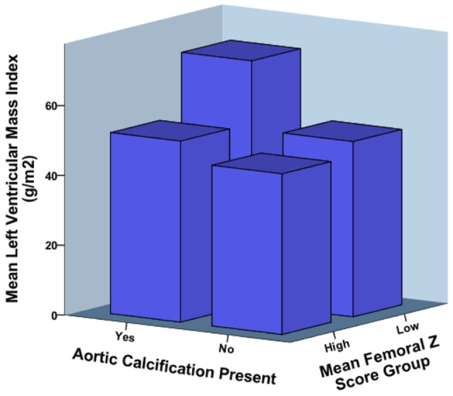
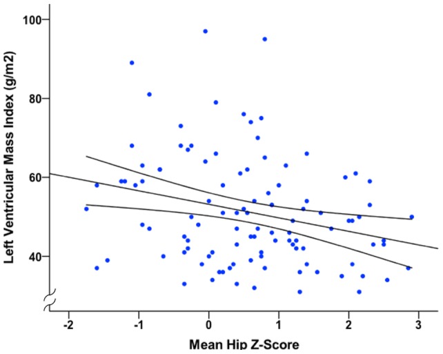
A total of 120 patients were recruited (54% male, mean age 55 ± 14 years, mean glomerular filtration rate 50 ± 13 ml/min/1.73 m(2)). Abdominal aortic calcification was assessed using lateral lumbar spine radiography and was present in 48%. Mean femoral Z-score measured using dual energy x-ray absorptiometry was 0.60 ± 1.06. Cardiovascular magnetic resonance imaging was used to determine left ventricular mass. One patient had left ventricular hypertrophy. Subjects with aortic calcification had higher left ventricular mass compared to those without (56 ± 16 vs. 48 ± 12 g/m(2), P = 0.002), as did patients with femoral Z-scores below zero (56 ± 15 vs. 49 ± 13 g/m(2), P = 0.01). In univariate analysis presence of aortic calcification correlated with left ventricular mass (r = 0.32, P = 0.001); mean femoral Z-score inversely correlated with left ventricular mass (r = -0.28, P = 0.004). In a multivariate regression model that included presence of aortic calcification, mean femoral Z-score, gender and 24-hour systolic blood pressure, 46% of the variability in left ventricular mass was explained (P<0.001).
In patients with stage 3 non-diabetic chronic kidney disease, lower mean femoral Z-score and presence of aortic calcification are independently associated with increased left ventricular mass. Further research exploring the pathophysiology that underlies these relationships is warranted.
血管钙化和骨密度降低在慢性肾脏病中很常见,与心血管风险增加有关。其机制尚不清楚。我们在一项横断面观察研究中评估了 3 期非糖尿病慢性肾脏病患者的血管钙化、股骨骨密度和左心室质量之间的关系。
共招募了 120 名患者(54%为男性,平均年龄 55 ± 14 岁,平均肾小球滤过率 50 ± 13 ml/min/1.73 m(2))。使用侧位腰椎 X 线摄影评估腹主动脉钙化,48%的患者存在钙化。使用双能 X 射线吸收法测量的股骨 Z 评分平均值为 0.60 ± 1.06。心血管磁共振成像用于确定左心室质量。1 名患者存在左心室肥厚。与无主动脉钙化的患者相比,有主动脉钙化的患者左心室质量更高(56 ± 16 与 48 ± 12 g/m(2),P = 0.002),股骨 Z 评分低于零的患者也是如此(56 ± 15 与 49 ± 13 g/m(2),P = 0.01)。在单变量分析中,主动脉钙化的存在与左心室质量相关(r = 0.32,P = 0.001);平均股骨 Z 评分与左心室质量呈负相关(r =-0.28,P = 0.004)。在包括主动脉钙化的存在、平均股骨 Z 评分、性别和 24 小时收缩压的多元回归模型中,左心室质量的可变性有 46%得到了解释(P<0.001)。
在 3 期非糖尿病慢性肾脏病患者中,平均股骨 Z 评分较低和主动脉钙化的存在与左心室质量增加独立相关。需要进一步研究探索这些关系背后的病理生理学。