• 文献检索
  • 文档翻译
  • 深度研究
  • 学术资讯
  • Suppr Zotero 插件Zotero 插件
  • 邀请有礼
  • 套餐&价格
  • 历史记录
应用&插件
Suppr Zotero 插件Zotero 插件浏览器插件Mac 客户端Windows 客户端微信小程序
定价
高级版会员购买积分包购买API积分包
服务
文献检索文档翻译深度研究API 文档MCP 服务
关于我们
关于 Suppr公司介绍联系我们用户协议隐私条款
关注我们

Suppr 超能文献

核心技术专利:CN118964589B侵权必究
粤ICP备2023148730 号-1Suppr @ 2026

文献检索

告别复杂PubMed语法,用中文像聊天一样搜索,搜遍4000万医学文献。AI智能推荐,让科研检索更轻松。

立即免费搜索

文件翻译

保留排版,准确专业,支持PDF/Word/PPT等文件格式,支持 12+语言互译。

免费翻译文档

深度研究

AI帮你快速写综述,25分钟生成高质量综述,智能提取关键信息,辅助科研写作。

立即免费体验

鼻窦提升术后施奈德膜下骨再生的生物学原理与生理学:一项针对14例行经牙槽嵴水动力超声空化鼻窦提升术(Intralift)患者的放射学研究

Biological Principles and Physiology of Bone Regeneration under the Schneiderian Membrane after Sinus Lift Surgery: A Radiological Study in 14 Patients Treated with the Transcrestal Hydrodynamic Ultrasonic Cavitational Sinus Lift (Intralift).

作者信息

Troedhan A, Kurrek A, Wainwright M

机构信息

Center for Facial Esthetics Vienna, Brauhausgasse 12-14, 1050 Vienna, Austria.

出版信息

Int J Dent. 2012;2012:576238. doi: 10.1155/2012/576238. Epub 2012 Jun 17.

DOI:10.1155/2012/576238
PMID:22754571
原文链接:https://pmc.ncbi.nlm.nih.gov/articles/PMC3382962/
Abstract

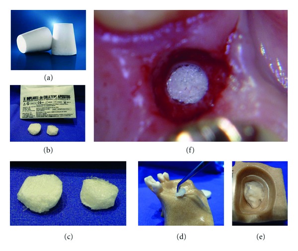
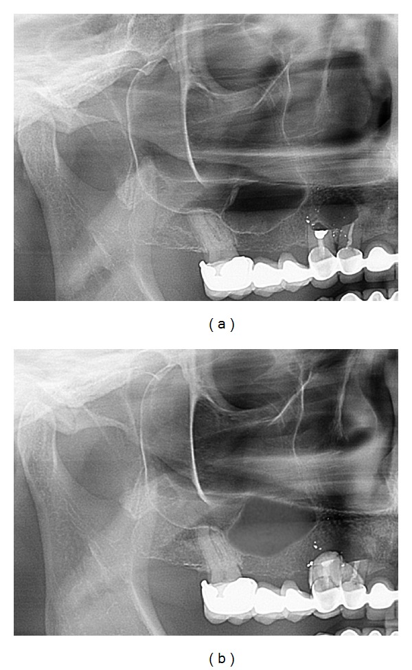
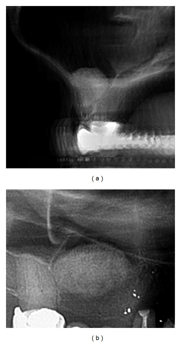
Introduction. Sinus lift procedures are a commonly accepted method of bone augmentation in the lateral maxilla with clinically good results. Nevertheless the role of the Schneiderian membrane in the bone-reformation process is discussed controversially. Aim of this study was to prove the key role of the sinus membrane in bone reformation in vivo. Material and Methods. 14 patients were treated with the minimal invasive tHUCSL-Intralift, and 2 ccm collagenous sponges were inserted subantrally and the calcification process followed up with CBCT scans 4 and 7 months after surgery. Results. An even and circular centripetal calcification under the sinus membrane and the antral floor was detected 4 months after surgery covering 30% of the entire augmentation width/height/depth at each wall. The calcification process was completed in the entire augmentation volume after 7 months. A loss of approximately 13% of absolute augmentation height was detected between the 4th and 7th month. Discussion. The results of this paper prove the key role of the sinus membrane as the main carrier of bone reformation after sinus lift procedures as multiple experimental studies suggested. Thus the importance of minimal invasive and rupture free sinuslift procedures is underlined and does not depend on the type of grafting material used.

摘要

引言。上颌窦提升术是上颌骨外侧常用的骨增量方法,临床效果良好。然而,关于施奈德膜在骨改建过程中的作用存在争议。本研究的目的是在体内证实上颌窦膜在骨改建中的关键作用。材料与方法。14例患者接受了微创经鼻上颌窦内提升术(tHUCSL-Intralift),将2立方厘米的胶原海绵植入上颌窦底,并在术后4个月和7个月通过锥形束计算机断层扫描(CBCT)追踪钙化过程。结果。术后4个月,在上颌窦膜和窦底下方检测到均匀的向心性钙化,覆盖每壁整个增量宽度/高度/深度的30%。钙化过程在7个月后在整个增量区域内完成。在第4个月至第7个月期间,检测到绝对增量高度损失约13%。讨论。本文结果证实了上颌窦膜作为上颌窦提升术后骨改建主要载体的关键作用,正如多项实验研究所表明的那样。因此,强调了微创且无破裂的上颌窦提升术的重要性,且其不依赖于所用移植材料的类型。

https://cdn.ncbi.nlm.nih.gov/pmc/blobs/8937/3382962/5be2f6cd5839/IJD2012-576238.020.jpg
https://cdn.ncbi.nlm.nih.gov/pmc/blobs/8937/3382962/0cd006b9b2d7/IJD2012-576238.001.jpg
https://cdn.ncbi.nlm.nih.gov/pmc/blobs/8937/3382962/466239c5a198/IJD2012-576238.002.jpg
https://cdn.ncbi.nlm.nih.gov/pmc/blobs/8937/3382962/ba0836ad1622/IJD2012-576238.003.jpg
https://cdn.ncbi.nlm.nih.gov/pmc/blobs/8937/3382962/c0ba60b526d6/IJD2012-576238.004.jpg
https://cdn.ncbi.nlm.nih.gov/pmc/blobs/8937/3382962/2da395cb70c5/IJD2012-576238.005.jpg
https://cdn.ncbi.nlm.nih.gov/pmc/blobs/8937/3382962/199985f8853a/IJD2012-576238.006.jpg
https://cdn.ncbi.nlm.nih.gov/pmc/blobs/8937/3382962/184944c8a77c/IJD2012-576238.007.jpg
https://cdn.ncbi.nlm.nih.gov/pmc/blobs/8937/3382962/dd2020ad7102/IJD2012-576238.008.jpg
https://cdn.ncbi.nlm.nih.gov/pmc/blobs/8937/3382962/ac8af257d448/IJD2012-576238.009.jpg
https://cdn.ncbi.nlm.nih.gov/pmc/blobs/8937/3382962/568fc48d1040/IJD2012-576238.010.jpg
https://cdn.ncbi.nlm.nih.gov/pmc/blobs/8937/3382962/d6eca1051be1/IJD2012-576238.011.jpg
https://cdn.ncbi.nlm.nih.gov/pmc/blobs/8937/3382962/2e0a3c22045b/IJD2012-576238.012.jpg
https://cdn.ncbi.nlm.nih.gov/pmc/blobs/8937/3382962/cdec10516086/IJD2012-576238.013.jpg
https://cdn.ncbi.nlm.nih.gov/pmc/blobs/8937/3382962/d243ecb7dc0e/IJD2012-576238.014.jpg
https://cdn.ncbi.nlm.nih.gov/pmc/blobs/8937/3382962/17e346548f52/IJD2012-576238.015.jpg
https://cdn.ncbi.nlm.nih.gov/pmc/blobs/8937/3382962/7775633bd306/IJD2012-576238.016.jpg
https://cdn.ncbi.nlm.nih.gov/pmc/blobs/8937/3382962/2156d08f5d37/IJD2012-576238.017.jpg
https://cdn.ncbi.nlm.nih.gov/pmc/blobs/8937/3382962/a12d6dd3668a/IJD2012-576238.018.jpg
https://cdn.ncbi.nlm.nih.gov/pmc/blobs/8937/3382962/fc06f08d2773/IJD2012-576238.019.jpg
https://cdn.ncbi.nlm.nih.gov/pmc/blobs/8937/3382962/5be2f6cd5839/IJD2012-576238.020.jpg

相似文献

1
Biological Principles and Physiology of Bone Regeneration under the Schneiderian Membrane after Sinus Lift Surgery: A Radiological Study in 14 Patients Treated with the Transcrestal Hydrodynamic Ultrasonic Cavitational Sinus Lift (Intralift).鼻窦提升术后施奈德膜下骨再生的生物学原理与生理学:一项针对14例行经牙槽嵴水动力超声空化鼻窦提升术(Intralift)患者的放射学研究
Int J Dent. 2012;2012:576238. doi: 10.1155/2012/576238. Epub 2012 Jun 17.
2
Comparison of Conventional Transcrestal Sinus Lift and Ultrasound-Enhanced Transcrestal Hydrodynamic Cavitational Sinus Lift for the Filling of Subantral Space: A Human Cadaver Study.传统经牙槽嵴上颌窦提升术与超声增强经牙槽嵴流体动力空化上颌窦提升术填充上颌窦底间隙的比较:一项人体尸体研究。
J Oral Implantol. 2015 Dec;41(6):657-61. doi: 10.1563/aaid-joi-D-14-00038. Epub 2014 Sep 18.
3
Schneiderian membrane detachment using transcrestal hydrodynamic ultrasonic cavitational sinus lift: a human cadaver head study and histologic analysis.
J Oral Maxillofac Surg. 2014 Aug;72(8):1503.e1-10. doi: 10.1016/j.joms.2014.02.021. Epub 2014 Feb 19.
4
Volumetric changes of grafted volumes and the Schneiderian membrane after transcrestal and lateral sinus floor elevation procedures: A clinical, pilot study.经牙槽嵴和外侧窦底提升术后移植物体积和黏膜体积的变化:一项临床初步研究。
J Clin Periodontol. 2017 Jun;44(6):660-671. doi: 10.1111/jcpe.12728. Epub 2017 May 16.
5
Effect of Schneiderian membrane perforation on sinus lift graft outcome using two different donor sites: a retrospective study of 105 maxillary sinus elevation procedures.使用两个不同供区时筛窦黏膜穿孔对上颌窦提升植骨结果的影响:一项对105例上颌窦提升手术的回顾性研究
GMS Interdiscip Plast Reconstr Surg DGPW. 2016 Mar 2;5:Doc11. doi: 10.3205/iprs000090. eCollection 2016.
6
Radiological sinus lift: a new minimally invasive CT-guided procedure for maxillary sinus floor elevation in implant dentistry.放射学引导下的上颌窦提升术:种植牙领域中一种新型的微创CT引导下的上颌窦底提升手术。
Clin Oral Implants Res. 2016 Mar;27(3):341-7. doi: 10.1111/clr.12549. Epub 2015 Jan 13.
7
The influence of sinus membrane thickness upon membrane perforation during transcrestal sinus lift procedure.经牙槽嵴上颌窦提升术中窦膜厚度对窦膜穿孔的影响。
Clin Oral Implants Res. 2015 Oct;26(10):1158-64. doi: 10.1111/clr.12429. Epub 2014 May 29.
8
Sinus floor augmentation with simultaneous implant placement using Choukroun's platelet-rich fibrin as the sole grafting material: a radiologic and histologic study at 6 months.应用 Choukroun 富血小板纤维蛋白作为唯一植骨材料同期进行种植体植入的牙槽嵴增高术:6 个月时的放射学和组织学研究。
J Periodontol. 2009 Dec;80(12):2056-64. doi: 10.1902/jop.2009.090252.
9
Histological and histomorphometric study using an ultrasonic crestal sinus grafting procedure. A multicenter case study.使用超声嵴顶窦底提升术的组织学和组织形态计量学研究。一项多中心病例研究。
Med Oral Patol Oral Cir Bucal. 2016 May 1;21(3):e367-73. doi: 10.4317/medoral.20994.
10
Rupture length of the sinus membrane after 1.2 mm puncture and surgical sinus elevation: an experimental animal cadaver study.1.2毫米穿刺及手术性上颌窦提升术后上颌窦膜破裂长度:一项实验动物尸体研究
Oral Surg Oral Med Oral Pathol Oral Radiol Endod. 2011 Nov;112(5):568-72. doi: 10.1016/j.tripleo.2010.11.014. Epub 2011 Feb 16.

引用本文的文献

1
Osseodensification versus piezoelectric internal sinus elevation (PISE) technique in delayed implant placement (a randomized controlled clinical trial).骨密度增强与压电内窦提升(PISE)技术在延迟种植体植入中的应用(一项随机对照临床试验)。
BMC Oral Health. 2024 Oct 28;24(1):1306. doi: 10.1186/s12903-024-04964-6.
2
Predictors of peri-implant bone remodeling outcomes after the osteotome sinus floor elevation: a retrospective study.经骨凿上颌窦底提升术后种植体周围骨重塑结局的预测因素:一项回顾性研究。
BMC Oral Health. 2022 Dec 20;22(1):622. doi: 10.1186/s12903-022-02592-6.
3
Bone Graft Packing and Its Association with Bone Regeneration in Maxillary Sinus Floor Augmentations: Histomorphometric Analysis of Human Biopsies.

本文引用的文献

1
Hydrodynamic ultrasonic maxillary sinus lift: review of a new technique and presentation of a clinical case.水动力超声上颌窦提升术:一种新技术的综述及临床病例报告。
Med Oral Patol Oral Cir Bucal. 2012 Mar 1;17(2):e271-5. doi: 10.4317/medoral.17430.
2
Rupture length of the sinus membrane after 1.2 mm puncture and surgical sinus elevation: an experimental animal cadaver study.1.2毫米穿刺及手术性上颌窦提升术后上颌窦膜破裂长度:一项实验动物尸体研究
Oral Surg Oral Med Oral Pathol Oral Radiol Endod. 2011 Nov;112(5):568-72. doi: 10.1016/j.tripleo.2010.11.014. Epub 2011 Feb 16.
3
The innate osteogenic potential of the maxillary sinus (Schneiderian) membrane: an ectopic tissue transplant model simulating sinus lifting.
骨移植填充及其在上颌窦底提升术中与骨再生的关系:人体活检组织形态计量学分析
Biology (Basel). 2022 Sep 30;11(10):1431. doi: 10.3390/biology11101431.
4
Management of Schneiderian Membrane Perforations during Sinus Augmentation Procedures: A Preliminary Comparison of Two Different Approaches.鼻窦增高术中施耐德膜穿孔的处理:两种不同方法的初步比较
J Clin Med. 2019 Sep 19;8(9):1491. doi: 10.3390/jcm8091491.
5
Histological and histomorphometric study using an ultrasonic crestal sinus grafting procedure. A multicenter case study.使用超声嵴顶窦底提升术的组织学和组织形态计量学研究。一项多中心病例研究。
Med Oral Patol Oral Cir Bucal. 2016 May 1;21(3):e367-73. doi: 10.4317/medoral.20994.
6
Biomechanical Stability of Dental Implants in Augmented Maxillary Sites: Results of a Randomized Clinical Study with Four Different Biomaterials and PRF and a Biological View on Guided Bone Regeneration.上颌骨增量部位种植体的生物力学稳定性:一项关于四种不同生物材料和富血小板纤维蛋白的随机临床研究结果及引导骨再生的生物学观点
Biomed Res Int. 2015;2015:850340. doi: 10.1155/2015/850340. Epub 2015 Apr 12.
7
Primary implant stability in augmented sinuslift-sites after completed bone regeneration: a randomized controlled clinical study comparing four subantrally inserted biomaterials.骨再生完成后上颌窦提升位点的种植体初期稳定性:一项比较四种经窦底植入生物材料的随机对照临床研究
Sci Rep. 2014 Jul 30;4:5877. doi: 10.1038/srep05877.
8
Graft-free sinus augmentation procedure: a literature review.无植骨窦底提升术:文献综述
J Oral Maxillofac Res. 2014 Apr 1;5(1):e1. doi: 10.5037/jomr.2014.5101. eCollection 2014 Jan.
上颌窦(施氏)膜的固有成骨潜能:模拟窦提升的异位组织移植模型。
Int J Oral Maxillofac Surg. 2010 Aug;39(8):793-801. doi: 10.1016/j.ijom.2010.03.009. Epub 2010 Apr 22.
4
Hydrodynamic ultrasonic sinus floor elevation--an experimental study in sheep.
J Oral Maxillofac Surg. 2010 May;68(5):1125-30. doi: 10.1016/j.joms.2009.12.014. Epub 2010 Mar 4.
5
The Schneiderian membrane contains osteoprogenitor cells: in vivo and in vitro study.筛骨膜含有骨祖细胞:体内和体外研究。
Calcif Tissue Int. 2009 Feb;84(2):138-45. doi: 10.1007/s00223-008-9202-x. Epub 2008 Dec 9.
6
[Ultrasonic bone cutting in oral surgery: a review of 60 cases].[口腔外科中的超声骨切割:60例病例回顾]
Ultraschall Med. 2008 Feb;29(1):66-71. doi: 10.1055/s-2007-963507.
7
De novo bone induction by recombinant human bone morphogenetic protein-2 (rhBMP-2) in maxillary sinus floor augmentation.重组人骨形态发生蛋白-2(rhBMP-2)在上颌窦底提升术中的新生骨诱导作用。
J Oral Maxillofac Surg. 2005 Dec;63(12):1693-707. doi: 10.1016/j.joms.2005.08.018.
8
Osteogenic capacities of periost grafts, periost flaps and prefabricated periosteal flaps: experimental study.骨膜移植、骨膜瓣及预制骨膜瓣的成骨能力:实验研究
J Craniofac Surg. 2005 Jul;16(4):594-600. doi: 10.1097/01.scs.0000168773.71356.62.
9
Bone reformation with sinus membrane elevation: a new surgical technique for maxillary sinus floor augmentation.伴窦膜提升的骨改建:一种上颌窦底提升的新手术技术
Clin Implant Dent Relat Res. 2004;6(3):165-73.
10
Bone induction capacity of the periosteum and neonatal dura in the setting of the rat zygomatic arch fracture model.大鼠颧弓骨折模型中骨膜和新生硬脑膜的骨诱导能力。
Arch Facial Plast Surg. 2003 Jul-Aug;5(4):301-8. doi: 10.1001/archfaci.5.4.301.