REPAIR-lab, Institute of Pathology, University Medical Center of the Johannes Gutenberg University, Langenbeckstraße 1, Mainz, 55101, Germany.
Part Fibre Toxicol. 2012 Jul 3;9:23. doi: 10.1186/1743-8977-9-23.
The use of gold nanoparticles (AuNPs) for diagnostic applications and for drug and gene-delivery is currently under intensive investigation. For such applications, biocompatibility and the absence of cytotoxicity of AuNPs is essential. Although generally considered as highly biocompatible, previous in vitro studies have shown that cytotoxicity of AuNPs in certain human epithelial cells was observed. In particular, the degree of purification of AuNPs (presence of sodium citrate residues on the particles) was shown to affect the proliferation and induce cytotoxicity in these cells. To expand these studies, we have examined if the effects are related to nanoparticle size (10, 11 nm, 25 nm), to the presence of sodium citrate on the particles' surface or they are due to a varying degree of internalization of the AuNPs. Since two cell types are present in the major barriers to the outside in the human body, we have also included endothelial cells from the vasculature and blood brain barrier.
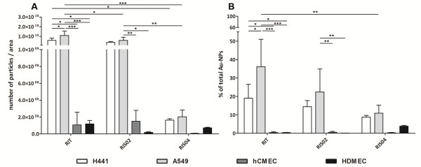
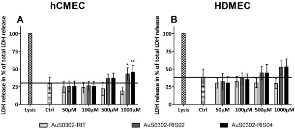
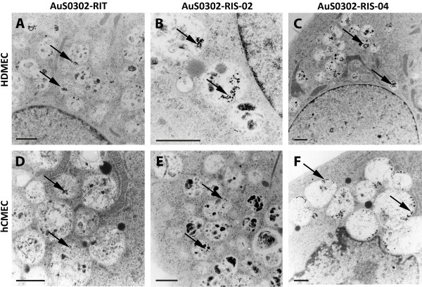
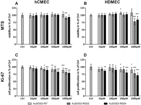
Transmission electron microscopy demonstrates that the internalized gold nanoparticles are located within vesicles. Increased cytotoxicity was observed after exposure to AuNPs and was found to be concentration-dependent. In addition, cell viability and the proliferation of both endothelial cells decreased after exposure to gold nanoparticles, especially at high concentrations. Moreover, in contrast to the size of the particles (10 nm, 11 nm, 25 nm), the presence of sodium citrate on the nanoparticle surface appeared to enhance these effects. The effects on microvascular endothelial cells from blood vessels were slightly enhanced compared to the effects on brain-derived endothelial cells. A quantification of AuNPs within cells by ICP-AES showed that epithelial cells internalized a higher quantity of AuNPs compared to endothelial cells and that the quantity of uptake is not correlated with the amount of sodium citrate on the nanoparticles' surface.
In conclusion the higher amount of citrate on the particle surface resulted in a higher impairment of cell viability, but did not enhance or reduce the uptake behavior in endothelial or epithelial cells. In addition, epithelial and endothelial cells exhibited different uptake behaviors for citrate-stabilized gold nanoparticles, which might be related to different interactions occurring at the nanoparticle-cell-surface interface. The different uptake in epithelial cells might explain the higher reduction of proliferation of these cells after exposure to AuNPs treatment although more detailed investigations are necessary to determine subcellular events. Nevertheless an extrinsic effect of sodium-citrate stabilized particles could not be excluded. Thus, the amount of sodium citrate should be reduced to a level on which the stability of the particles and the safety for biomedical applications are guaranteed.
目前,金纳米粒子(AuNPs)在诊断应用以及药物和基因传递方面的应用正受到广泛关注。对于此类应用,AuNPs 的生物相容性和无细胞毒性至关重要。尽管通常被认为具有高度生物相容性,但先前的体外研究表明,在某些人类上皮细胞中观察到 AuNPs 的细胞毒性。特别是,AuNPs 的纯化程度(颗粒表面存在柠檬酸钠残留物)被证明会影响这些细胞的增殖并诱导细胞毒性。为了扩展这些研究,我们检查了这些效应是否与纳米颗粒大小(10、11nm、25nm)、颗粒表面上存在柠檬酸钠或由于 AuNPs 内化程度不同有关。由于人体外部的主要屏障中存在两种细胞类型,我们还包括了血管内皮细胞和血脑屏障。
透射电子显微镜显示,内化的金纳米粒子位于囊泡内。暴露于 AuNPs 后观察到细胞毒性增加,且呈浓度依赖性。此外,暴露于金纳米粒子后,两种内皮细胞的细胞活力和增殖均下降,尤其是在高浓度下。此外,与颗粒大小(10nm、11nm、25nm)相比,颗粒表面上存在柠檬酸钠似乎增强了这些效应。与脑源性内皮细胞相比,血管内皮细胞的微血管内皮细胞的效应略有增强。通过 ICP-AES 对细胞内 AuNPs 的定量显示,上皮细胞内化的 AuNPs 量高于内皮细胞,并且摄取量与纳米颗粒表面上的柠檬酸钠量无关。
总之,颗粒表面上更多的柠檬酸导致细胞活力的更大损害,但并未增强或减少内皮细胞或上皮细胞的摄取行为。此外,上皮细胞和内皮细胞对柠檬酸稳定的金纳米粒子表现出不同的摄取行为,这可能与纳米颗粒-细胞表面界面处发生的不同相互作用有关。尽管需要更详细的研究来确定亚细胞事件,但上皮细胞增殖减少可能是由于暴露于 AuNP 处理后这些细胞的摄取不同所致。然而,不能排除柠檬酸稳定的颗粒的外在影响。因此,应该将柠檬酸钠的量减少到保证颗粒稳定性和生物医学应用安全性的水平。