Department of Neurology and Neurological Sciences, Stanford University School of Medicine, Stanford, CA 94305, USA.
Neurobiol Dis. 2012 Dec;48(3):429-38. doi: 10.1016/j.nbd.2012.06.019. Epub 2012 Jul 2.
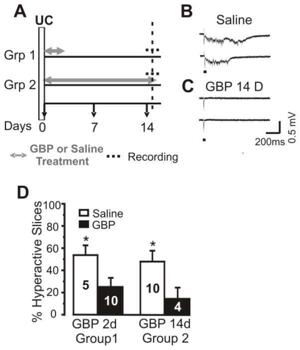
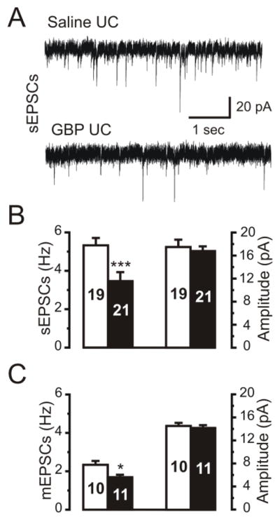
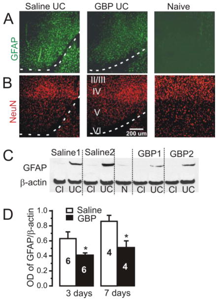
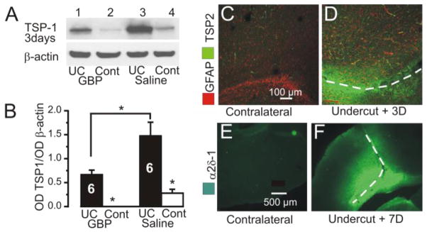
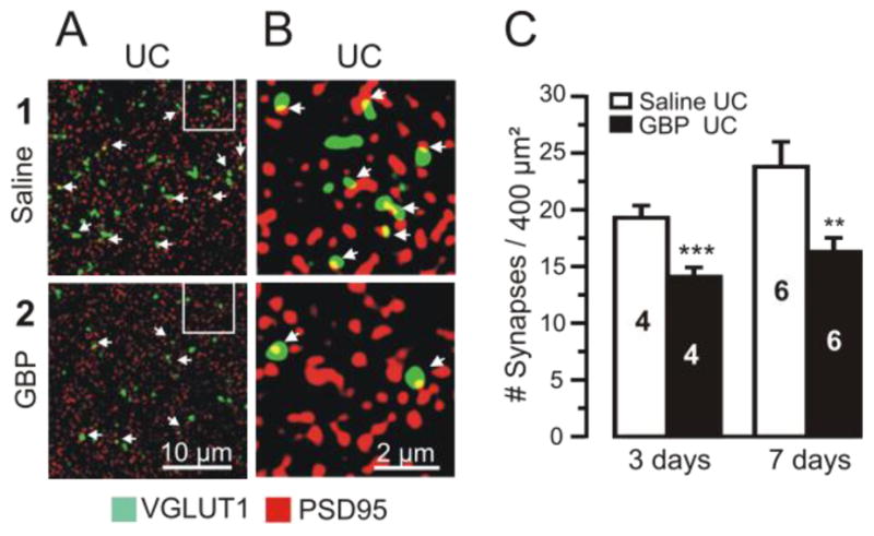
Gabapentin (GBP) is an anticonvulsant that acts at the α2δ-1 submit of the L-type calcium channel. It is recently reported that GBP is a potent inhibitor of thrombospondin (TSP)-induced excitatory synapse formation in vitro and in vivo. Here we studied effects of chronic GBP administration on epileptogenesis in the partial cortical isolation ("undercut") model of posttraumatic epilepsy, in which abnormal axonal sprouting and aberrant synaptogenesis contribute to occurrence of epileptiform discharges. Results showed that 1) the incidence of evoked epileptiform discharges in undercut cortical slices studied 1 day or ~2 weeks after the last GBP dose, was significantly reduced by GBP treatments, beginning on the day of injury; 2) the expression of GFAP and TSP1 protein, as well as the number of FJC stained cells was decreased in GBP treated undercut animals; 3) in vivo GBP treatment of rats with undercuts for 3 or 7 days decreased the density of vGlut1-PSD95 close appositions (presumed synapses) in comparison to saline treated controls with similar lesions;4) the electrophysiological data are compatible with the above anatomical changes, showing decreases in mEPSC and sEPSC frequency in the GBP treated animals. These results indicate that chronic administration of GBP after cortical injury is antiepileptogenic in the undercut model of post-traumatic epilepsy, perhaps by both neuroprotective actions and decreases in excitatory synapse formation. The findings may suggest the potential use of GBP as an antiepileptogenic agent following traumatic brain injury.
加巴喷丁(GBP)是一种抗惊厥药,作用于 L 型钙通道的α2δ-1 亚基。最近有报道称,GBP 是一种有效的血小板反应蛋白(TSP)诱导的体外和体内兴奋性突触形成抑制剂。在这里,我们研究了慢性 GBP 给药对创伤后癫痫部分皮质隔离(“切迹”)模型中癫痫发生的影响,其中异常轴突发芽和异常突触发生有助于癫痫样放电的发生。结果表明:1)在最后一次 GBP 剂量后 1 天或约 2 周研究的切迹皮质切片中,诱发癫痫样放电的发生率通过 GBP 治疗显著降低,起始于损伤当天;2)GBP 处理的切迹动物中 GFAP 和 TSP1 蛋白的表达以及 FJC 染色细胞的数量减少;3)在体内 GBP 治疗切迹大鼠 3 或 7 天与盐水处理的具有相似损伤的对照组相比,降低了 vGlut1-PSD95 紧密贴合(假定的突触)的密度;4)电生理数据与上述解剖变化一致,显示 GBP 处理动物的 mEPSC 和 sEPSC 频率降低。这些结果表明,皮质损伤后慢性 GBP 给药在创伤后癫痫的切迹模型中具有抗癫痫发生作用,这可能是通过神经保护作用和减少兴奋性突触形成来实现的。这些发现可能表明 GBP 作为创伤性脑损伤后抗癫痫发生剂的潜在用途。