Department of Radiation Oncology, Wake Forest University School of Medicine, Winston-Salem, NC 27157, USA.
J Appl Clin Med Phys. 2012 Jul 5;13(4):3613. doi: 10.1120/jacmp.v13i4.3613.
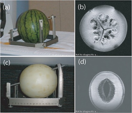
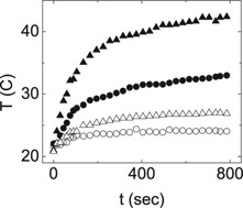
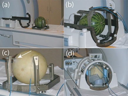
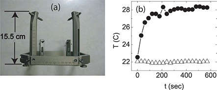
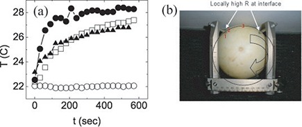
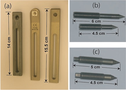
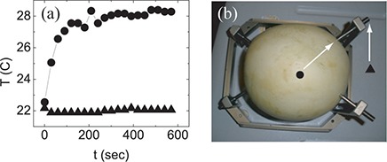
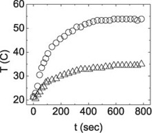
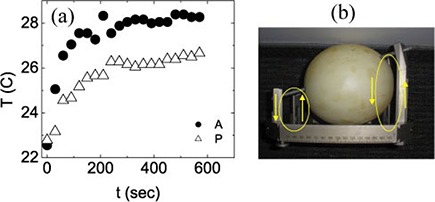
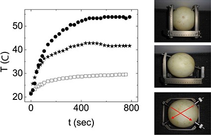
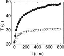
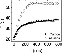
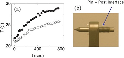
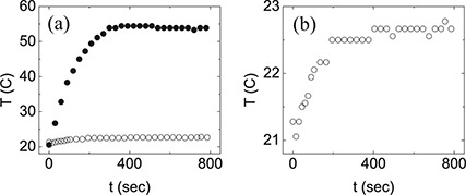
Magnetic resonance imaging (MRI) is regularly used for stereotactic imaging of Gamma Knife (GK) radiosurgery patients for GK treatment planning. MRI-induced thermal injuries have occurred and been reported for GK patients with attached metallic headframes. Depending on the specific MR imaging and headframe conditions, a skin injury from MRI-induced heating can potentially occur where the four headframe screws contact the skin surface of the patient's head. Higher MR field strength has a greater heating potential. Two primary heating mechanisms, electromagnetic induction and the antenna effect, are possible. In this study, MRI-induced heating from a 3T clinical MRI scanner was investigated for stereotactic headframes used in gamma radiosurgery and neurosurgery. Using melons as head phantoms, optical thermometers were used to characterize the temperature profile at various points of the melon headframe composite as a function of two 3T MR pulse sequence protocols. Different combinations of GK radiosurgery headframe post and screw designs were tested to determine best and worst combinations for MRI-induced heating. Temperature increases were measured for all pulse sequences tested, indicating that the potential exists for MRI-induced skin heating and burns at the headframe attachment site. This heating originates with electromagnetic induction caused by the RF fields inducing current in a loop formed by the headframe, mounting screws, and the region of the patient's head located between any of the two screws. This induced current is then resistively dissipated, with the regions of highest resistance, located at the headframe screw-patient head interface, experiencing the most heating. Significant heating can be prevented by replacing the metallic threads holding the screw with electrically insulated nuts, which is the heating prevention and patient safety recommendation of the GK manufacturer. Our results confirm that the manufacturer's recommendation to use insulating nuts reduces the induced currents in the headframe nearly to zero, effectively preventing heating and minimizing the likelihood of thermal injury.
磁共振成像(MRI)常用于伽玛刀(GK)放射外科患者的立体定向成像,以进行 GK 治疗计划。已经发生并报道了与附着的金属头架的 GK 患者的 MRI 诱导热损伤。根据特定的磁共振成像和头架条件,在四个头架螺钉接触患者头部皮肤表面的地方,可能会发生由 MRI 诱导加热引起的皮肤损伤。更高的磁共振场强具有更大的加热潜力。可能存在两种主要的加热机制,即电磁感应和天线效应。在这项研究中,研究了用于伽玛放射外科和神经外科的立体定向头架在 3T 临床磁共振扫描仪中的 MRI 诱导加热。使用甜瓜作为头部模型,使用光学温度计来描绘甜瓜头架复合材料各个点的温度分布,作为两种 3T MR 脉冲序列方案的函数。测试了不同的 GK 放射外科头架柱和螺钉设计组合,以确定最适合和最不适合 MRI 诱导加热的组合。测试了所有脉冲序列,均测量到温度升高,表明存在头架附件部位 MRI 诱导皮肤加热和灼伤的潜在风险。这种加热源自射频场在头架、安装螺钉和位于两个螺钉之间的患者头部区域形成的环路中感应电流引起的电磁感应。然后,感应电流会以电阻的形式耗散,电阻最高的区域位于头架螺钉-患者头部界面处,因此会经历最强烈的加热。通过用电气绝缘螺母代替固定螺钉的金属螺纹,可以防止这种加热,这是 GK 制造商的加热预防和患者安全建议。我们的结果证实,制造商建议使用绝缘螺母可使头架中的感应电流几乎降至零,从而有效防止加热并最大程度地降低热损伤的可能性。