Laboratory of Chronopharmacology, Institute of Biosciences, University of São Paulo, São Paulo, SP, Brazil.
PLoS One. 2012;7(7):e40142. doi: 10.1371/journal.pone.0040142. Epub 2012 Jul 2.
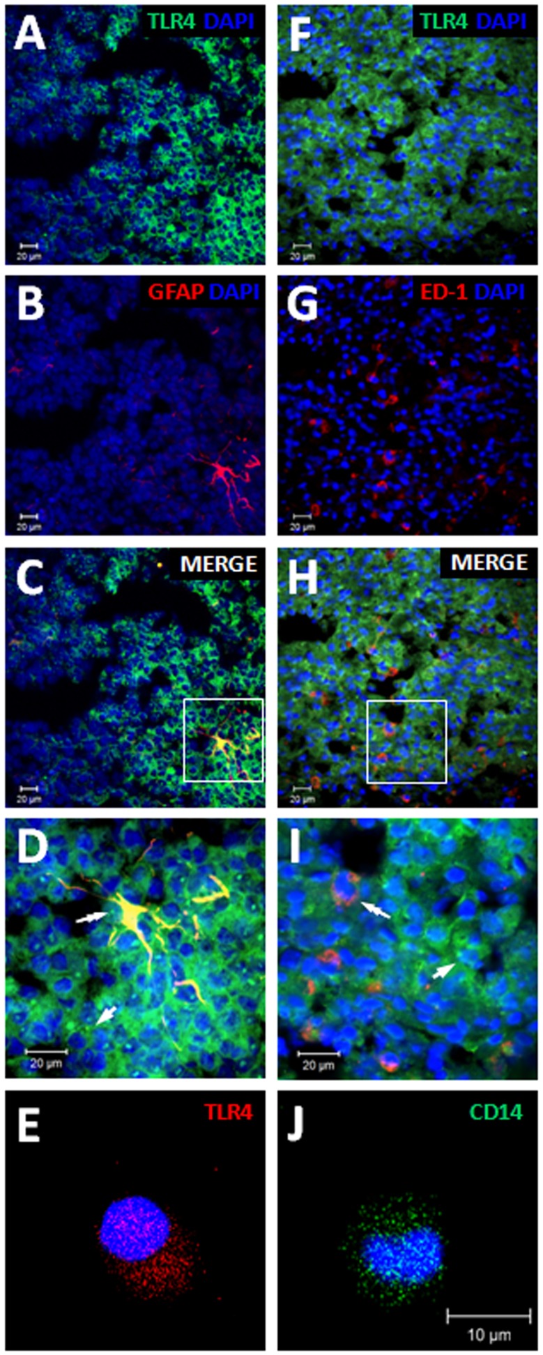
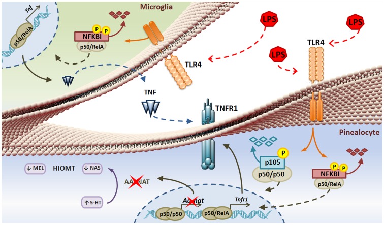
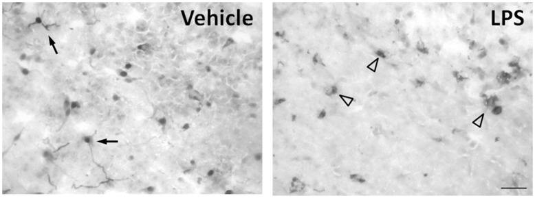
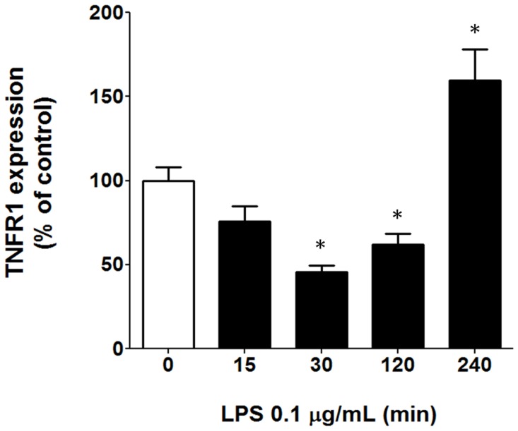
The pineal gland, a circumventricular organ, plays an integrative role in defense responses. The injury-induced suppression of the pineal gland hormone, melatonin, which is triggered by darkness, allows the mounting of innate immune responses. We have previously shown that cultured pineal glands, which express toll-like receptor 4 (TLR4) and tumor necrosis factor receptor 1 (TNFR1), produce TNF when challenged with lipopolysaccharide (LPS). Here our aim was to evaluate which cells present in the pineal gland, astrocytes, microglia or pinealocytes produced TNF, in order to understand the interaction between pineal activity, melatonin production and immune function. Cultured pineal glands or pinealocytes were stimulated with LPS. TNF content was measured using an enzyme-linked immunosorbent assay. TLR4 and TNFR1 expression were analyzed by confocal microscopy. Microglial morphology was analyzed by immunohistochemistry. In the present study, we show that although the main cell types of the pineal gland (pinealocytes, astrocytes and microglia) express TLR4, the production of TNF induced by LPS is mediated by microglia. This effect is due to activation of the nuclear factor kappa B (NF-kB) pathway. In addition, we observed that LPS activates microglia and modulates the expression of TNFR1 in pinealocytes. As TNF has been shown to amplify and prolong inflammatory responses, its production by pineal microglia suggests a glia-pinealocyte network that regulates melatonin output. The current study demonstrates the molecular and cellular basis for understanding how melatonin synthesis is regulated during an innate immune response, thus our results reinforce the role of the pineal gland as sensor of immune status.
松果腺作为一个室周器官,在防御反应中发挥整合作用。由黑暗引发的松果腺激素褪黑素的损伤抑制,允许固有免疫反应的启动。我们之前已经表明,表达 Toll 样受体 4 (TLR4) 和肿瘤坏死因子受体 1 (TNFR1) 的培养的松果腺在受到脂多糖 (LPS) 挑战时会产生 TNF。在这里,我们的目的是评估在松果腺中,星形胶质细胞、小胶质细胞或松果体细胞产生 TNF 的是哪些细胞,以了解松果腺活性、褪黑素产生和免疫功能之间的相互作用。用 LPS 刺激培养的松果腺或松果体细胞。使用酶联免疫吸附测定法测量 TNF 含量。通过共聚焦显微镜分析 TLR4 和 TNFR1 的表达。通过免疫组织化学分析小胶质细胞形态。在本研究中,我们表明,尽管松果腺的主要细胞类型(松果体细胞、星形胶质细胞和小胶质细胞)表达 TLR4,但 LPS 诱导的 TNF 产生是由小胶质细胞介导的。这种作用是由于核因子 kappa B (NF-kB) 途径的激活。此外,我们观察到 LPS 激活小胶质细胞并调节松果体细胞中 TNFR1 的表达。由于 TNF 已被证明可放大和延长炎症反应,因此松果体小胶质细胞产生的 TNF 表明存在调节褪黑素输出的神经胶质-松果体细胞网络。本研究为理解在固有免疫反应期间褪黑素合成如何受到调节提供了分子和细胞基础,因此我们的结果加强了松果腺作为免疫状态传感器的作用。