• 文献检索
  • 文档翻译
  • 深度研究
  • 学术资讯
  • Suppr Zotero 插件Zotero 插件
  • 邀请有礼
  • 套餐&价格
  • 历史记录
应用&插件
Suppr Zotero 插件Zotero 插件浏览器插件Mac 客户端Windows 客户端微信小程序
定价
高级版会员购买积分包购买API积分包
服务
文献检索文档翻译深度研究API 文档MCP 服务
关于我们
关于 Suppr公司介绍联系我们用户协议隐私条款
关注我们

Suppr 超能文献

核心技术专利:CN118964589B侵权必究
粤ICP备2023148730 号-1Suppr @ 2026

文献检索

告别复杂PubMed语法,用中文像聊天一样搜索,搜遍4000万医学文献。AI智能推荐,让科研检索更轻松。

立即免费搜索

文件翻译

保留排版,准确专业,支持PDF/Word/PPT等文件格式,支持 12+语言互译。

免费翻译文档

深度研究

AI帮你快速写综述,25分钟生成高质量综述,智能提取关键信息,辅助科研写作。

立即免费体验

胰腺癌微环境分析:巨噬细胞浸润及生长因子表达的作用

Analysis of pancreatic cancer microenvironment: role of macrophage infiltrates and growth factors expression.

作者信息

Gardian Katarzyna, Janczewska Sława, Olszewski Waldemar L, Durlik Marek

机构信息

Mossakowski Medical Research Centre Polish Academy of Sciences, Department of Surgical Research and Transplantology 5 Pawinskiego Str, 02-106 Warsaw, POLAND.

出版信息

J Cancer. 2012;3:285-91. doi: 10.7150/jca.4537. Epub 2012 Jul 1.

DOI:10.7150/jca.4537
PMID:22773932
原文链接:https://pmc.ncbi.nlm.nih.gov/articles/PMC3390598/
Abstract

BACKGROUND

Research over the last twenty years has yielded much insight into pancreatic cancer biology, but it has neither improved diagnostics methods nor the way of treatment. The question remains as to what the critical deciding factor is in making pancreatic cancer such an aggressive disease.

METHODS

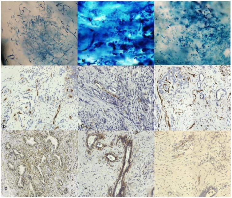
Pancreatic tumor tissue came from 36 patients. To assess lymphatic vessels color lymphangiography and immunohistochemistry were used. Activity of matrix metalloproteinases was studied with gel and in situ zymography. Expression of growth factors and infiltrating immune cells were investigated using immunohistochemistry.

RESULTS

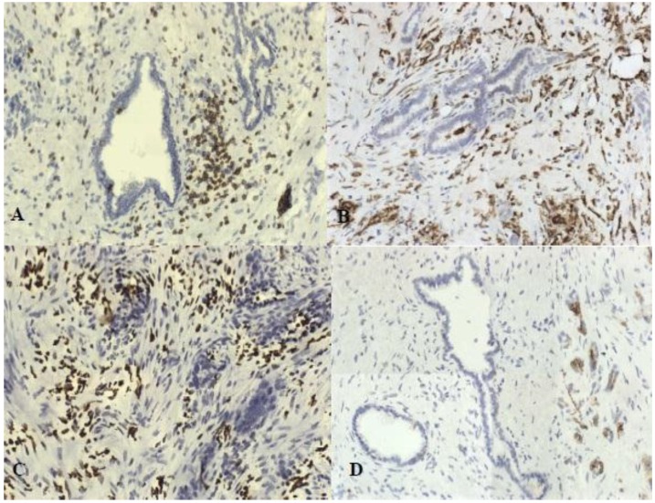
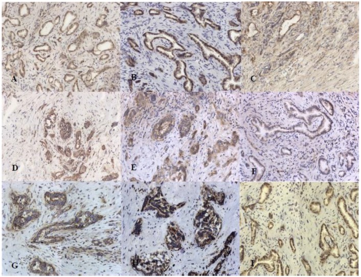
Our study revealed that the structures that correspond to lymphatic vessels were not observed in tumor center but only at the edge of the tumor. All studied growth factors were present in tumor tissue. We found that the difference in expression between G2 and G3 stage was statistically relevant in cases of c-Met receptor. Inflammatory cells were present around neoplastic glands and also strongly around nerves infiltrated by cancer cells. The number of infiltrating macrophages in tumor tissue was significantly higher in group with metastases to lymph nodes.

CONCLUSION

We showed two factors that influence pancreatic cancer progression and invasion: c-Met receptors and macrophages infiltrating tumor tissue. Based on our analysis, this indicates that epithelial-mesenchymal transition might be crucial in the progression of pancreatic cancer.

摘要

背景

过去二十年的研究对胰腺癌生物学有了深入了解,但既未改进诊断方法,也未改善治疗方式。胰腺癌为何如此具有侵袭性,关键决定因素究竟是什么,这一问题依然存在。

方法

36例患者的胰腺肿瘤组织用于研究。采用彩色淋巴管造影和免疫组化评估淋巴管。通过凝胶和原位酶谱分析研究基质金属蛋白酶的活性。利用免疫组化研究生长因子的表达及浸润免疫细胞。

结果

我们的研究表明,肿瘤中心未观察到与淋巴管相对应的结构,仅在肿瘤边缘发现。所有研究的生长因子均存在于肿瘤组织中。我们发现,在c-Met受体病例中,G2期和G3期之间的表达差异具有统计学意义。炎性细胞存在于肿瘤腺体周围,在癌细胞浸润的神经周围也大量存在。有淋巴结转移组的肿瘤组织中浸润巨噬细胞的数量显著更高。

结论

我们发现了影响胰腺癌进展和侵袭的两个因素:c-Met受体和浸润肿瘤组织的巨噬细胞。基于我们的分析,这表明上皮-间质转化可能在胰腺癌进展中起关键作用。

https://cdn.ncbi.nlm.nih.gov/pmc/blobs/a6fa/3390598/06d93c468bcd/jcav03p0285g03.jpg
https://cdn.ncbi.nlm.nih.gov/pmc/blobs/a6fa/3390598/02fdd405f7ca/jcav03p0285g01.jpg
https://cdn.ncbi.nlm.nih.gov/pmc/blobs/a6fa/3390598/0cfe52085a1f/jcav03p0285g02.jpg
https://cdn.ncbi.nlm.nih.gov/pmc/blobs/a6fa/3390598/06d93c468bcd/jcav03p0285g03.jpg
https://cdn.ncbi.nlm.nih.gov/pmc/blobs/a6fa/3390598/02fdd405f7ca/jcav03p0285g01.jpg
https://cdn.ncbi.nlm.nih.gov/pmc/blobs/a6fa/3390598/0cfe52085a1f/jcav03p0285g02.jpg
https://cdn.ncbi.nlm.nih.gov/pmc/blobs/a6fa/3390598/06d93c468bcd/jcav03p0285g03.jpg

相似文献

1
Analysis of pancreatic cancer microenvironment: role of macrophage infiltrates and growth factors expression.胰腺癌微环境分析:巨噬细胞浸润及生长因子表达的作用
J Cancer. 2012;3:285-91. doi: 10.7150/jca.4537. Epub 2012 Jul 1.
2
Microenvironment elements involved in the development of pancreatic cancer tumor.参与胰腺癌肿瘤发展的微环境元素。
Gastroenterol Res Pract. 2012;2012:585674. doi: 10.1155/2012/585674. Epub 2012 Dec 13.
3
M2-polarized tumor-associated macrophage infiltration of regional lymph nodes is associated with nodal lymphangiogenesis and occult nodal involvement in pN0 pancreatic cancer.区域淋巴结中 M2 极化的肿瘤相关巨噬细胞浸润与 pN0 胰腺癌中的淋巴结淋巴管生成和隐匿性淋巴结受累相关。
Pancreas. 2013 Jan;42(1):155-9. doi: 10.1097/MPA.0b013e318254f2d1.
4
Role of lymphangiogenesis and lymphangiogenic factors during pancreatic cancer progression and lymphatic spread.淋巴管生成及淋巴管生成因子在胰腺癌进展和淋巴转移过程中的作用
Int J Oncol. 2006 Apr;28(4):883-90.
5
Vascular endothelial growth factor-D induces lymphangiogenesis and lymphatic metastasis in models of ductal pancreatic cancer.血管内皮生长因子-D在胰腺导管癌模型中诱导淋巴管生成和淋巴转移。
Int J Oncol. 2005 Sep;27(3):669-79.
6
Tumor-associated macrophages induce capillary morphogenesis of lymphatic endothelial cells derived from human gastric cancer.肿瘤相关巨噬细胞诱导源自人胃癌的淋巴管内皮细胞发生毛细血管形态发生。
Cancer Sci. 2016 Aug;107(8):1101-9. doi: 10.1111/cas.12977. Epub 2016 Jun 22.
7
Analysis of tumor-induced lymphangiogenesis and lymphatic vessel invasion of pancreatic carcinoma in the peripheral nerve plexus.分析胰腺癌在周围神经丛中诱导的淋巴管生成和淋巴管浸润。
Cancer Sci. 2012 Oct;103(10):1756-63. doi: 10.1111/j.1349-7006.2012.02364.x. Epub 2012 Aug 27.
8
Protease-activated receptor 2 suppresses lymphangiogenesis and subsequent lymph node metastasis in a murine pancreatic cancer model.蛋白酶激活受体2在小鼠胰腺癌模型中抑制淋巴管生成及随后的淋巴结转移。
J Pathol. 2014 Nov;234(3):398-409. doi: 10.1002/path.4411. Epub 2014 Sep 12.
9
Soluble vascular endothelial growth factor receptor-3 suppresses lymphangiogenesis and lymphatic metastasis in bladder cancer.可溶性血管内皮生长因子受体-3 抑制膀胱癌中的淋巴管生成和淋巴转移。
Mol Cancer. 2011 Apr 11;10:36. doi: 10.1186/1476-4598-10-36.
10
Docetaxel facilitates lymphatic-tumor crosstalk to promote lymphangiogenesis and cancer progression.多西他赛促进淋巴管-肿瘤串扰以促进淋巴管生成和癌症进展。
BMC Cancer. 2018 Jul 6;18(1):718. doi: 10.1186/s12885-018-4619-8.

引用本文的文献

1
Effects of the superior mesenteric artery approach versus the no-touch approach during pancreatoduodenectomy on the mobilization of circulating tumour cells and clusters in pancreatic cancer (CETUPANC): randomized clinical trial.肠系膜上动脉入路与无接触入路在胰十二指肠切除术中对胰腺癌循环肿瘤细胞和簇转移的影响(CETUPANC):随机临床试验。
BJS Open. 2024 Oct 29;8(6). doi: 10.1093/bjsopen/zrae123.
2
Immune cells and checkpoints in pancreatic adenocarcinoma: Association with clinical and pathological characteristics.胰腺腺癌中的免疫细胞和检查点:与临床和病理特征的关联。
PLoS One. 2024 Jul 2;19(7):e0305648. doi: 10.1371/journal.pone.0305648. eCollection 2024.
3

本文引用的文献

1
Expression and prognostic value of circulating angiogenic cytokines in pancreatic cancer.循环血管生成细胞因子在胰腺癌中的表达及其预后价值。
BMC Cancer. 2011 Jul 5;11:286. doi: 10.1186/1471-2407-11-286.
2
Targeting the insulin growth factor pathway in gastrointestinal cancers.针对胃肠道癌症中的胰岛素样生长因子途径。
Oncology (Williston Park). 2011 May;25(6):518-26, 529.
3
Lack of functioning lymphatics and accumulation of tissue fluid/lymph in interstitial "lakes" in colon cancer tissue.结直肠癌组织中淋巴管功能缺失和组织液/淋巴在间质“湖泊”中积聚。
Roles of differently polarized macrophages in the initiation and progressionof pancreatic cancer.
不同极化状态的巨噬细胞在胰腺癌的发生发展中的作用。
Front Immunol. 2023 Aug 11;14:1237711. doi: 10.3389/fimmu.2023.1237711. eCollection 2023.
4
Perineural Invasion and Associated Pain Transmission in Pancreatic Cancer.胰腺癌中的神经周围浸润及相关疼痛传导
Cancers (Basel). 2021 Sep 13;13(18):4594. doi: 10.3390/cancers13184594.
5
Loss of activating transcription factor 3 prevents KRAS-mediated pancreatic cancer.激活转录因子 3 的缺失可预防 KRAS 介导的胰腺癌。
Oncogene. 2021 Apr;40(17):3118-3135. doi: 10.1038/s41388-021-01771-z. Epub 2021 Apr 16.
6
CXCL12 and Its Isoforms: Different Roles in Pancreatic Cancer?CXCL12 及其异构体:在胰腺癌中发挥不同作用?
J Oncol. 2019 Jun 2;2019:9681698. doi: 10.1155/2019/9681698. eCollection 2019.
7
Advances in Translational Research and Clinical Care in Pancreatic Cancer: Where Are We Headed?胰腺癌转化研究与临床护理的进展:我们将走向何方?
Gastroenterol Res Pract. 2019 Feb 4;2019:7690528. doi: 10.1155/2019/7690528. eCollection 2019.
8
Targeting Intramembrane Protein-Protein Interactions: Novel Therapeutic Strategy of Millions Years Old.靶向跨膜蛋白-蛋白相互作用:数百万年历史的新型治疗策略。
Adv Protein Chem Struct Biol. 2018;111:61-99. doi: 10.1016/bs.apcsb.2017.06.004. Epub 2017 Jul 24.
9
Extracellular Influences: Molecular Subclasses and the Microenvironment in Pancreatic Cancer.细胞外影响因素:胰腺癌的分子亚类与微环境
Cancers (Basel). 2018 Jan 27;10(2):34. doi: 10.3390/cancers10020034.
10
Novel TREM-1 Inhibitors Attenuate Tumor Growth and Prolong Survival in Experimental Pancreatic Cancer.新型 Trem-1 抑制剂可抑制实验性胰腺癌肿瘤生长和延长生存期。
Mol Pharm. 2017 Dec 4;14(12):4572-4582. doi: 10.1021/acs.molpharmaceut.7b00711. Epub 2017 Nov 13.
Lymphology. 2010 Dec;43(4):158-67.
4
Significance of lymphangiogenesis in primary tumor and draining lymph nodes during lymphatic metastasis of pancreatic head cancer.在胰头癌淋巴转移过程中,原发肿瘤和引流淋巴结淋巴管生成的意义。
J Surg Oncol. 2010 Dec 1;102(7):809-15. doi: 10.1002/jso.21744.
5
Macrophage diversity enhances tumor progression and metastasis.巨噬细胞多样性增强肿瘤的进展和转移。
Cell. 2010 Apr 2;141(1):39-51. doi: 10.1016/j.cell.2010.03.014.
6
Molecular mechanism of pancreatic cancer--understanding proliferation, invasion, and metastasis.胰腺癌的分子机制——了解增殖、浸润和转移。
Langenbecks Arch Surg. 2010 Apr;395(4):295-308. doi: 10.1007/s00423-010-0622-5. Epub 2010 Mar 18.
7
Lymphangiogenesis: Molecular mechanisms and future promise.淋巴管生成:分子机制与未来前景。
Cell. 2010 Feb 19;140(4):460-76. doi: 10.1016/j.cell.2010.01.045.
8
Macrophage matrix metalloproteinase-9 mediates epithelial-mesenchymal transition in vitro in murine renal tubular cells.巨噬细胞基质金属蛋白酶-9在体外介导小鼠肾小管细胞的上皮-间充质转化。
Am J Pathol. 2010 Mar;176(3):1256-70. doi: 10.2353/ajpath.2010.090188. Epub 2010 Jan 14.
9
Increased intratumoral lymphatic vessel density correlates with lymph node metastasis in early gastric carcinoma.肿瘤内淋巴管密度增加与早期胃癌淋巴结转移相关。
Ann Surg Oncol. 2010 Jan;17(1):73-80. doi: 10.1245/s10434-009-0707-y. Epub 2009 Sep 24.
10
Significance of M2-polarized tumor-associated macrophage in pancreatic cancer.M2 型肿瘤相关巨噬细胞在胰腺癌中的意义。
J Surg Res. 2011 May 15;167(2):e211-9. doi: 10.1016/j.jss.2009.05.026. Epub 2009 Jun 16.