Muldoon Timothy J, Burgess Sean A, Chen Brenda R, Ratner Désirée, Hillman Elizabeth M C
Biomed Opt Express. 2012 Jul 1;3(7):1701-12. doi: 10.1364/BOE.3.001701. Epub 2012 Jun 22.
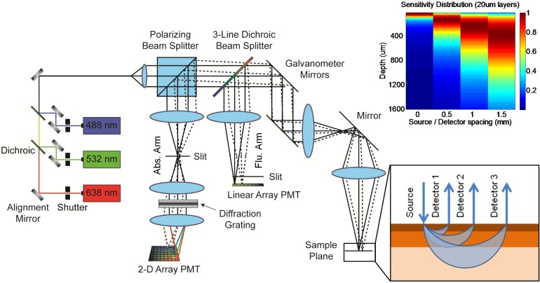
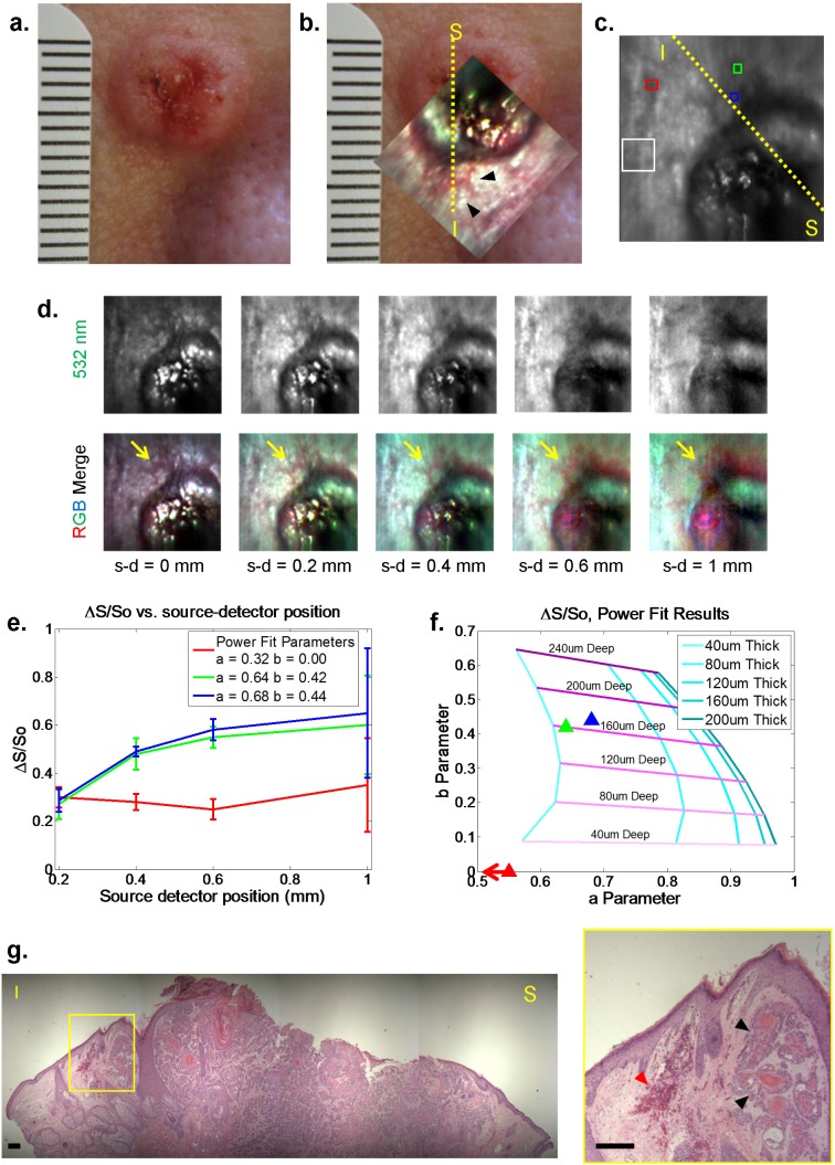
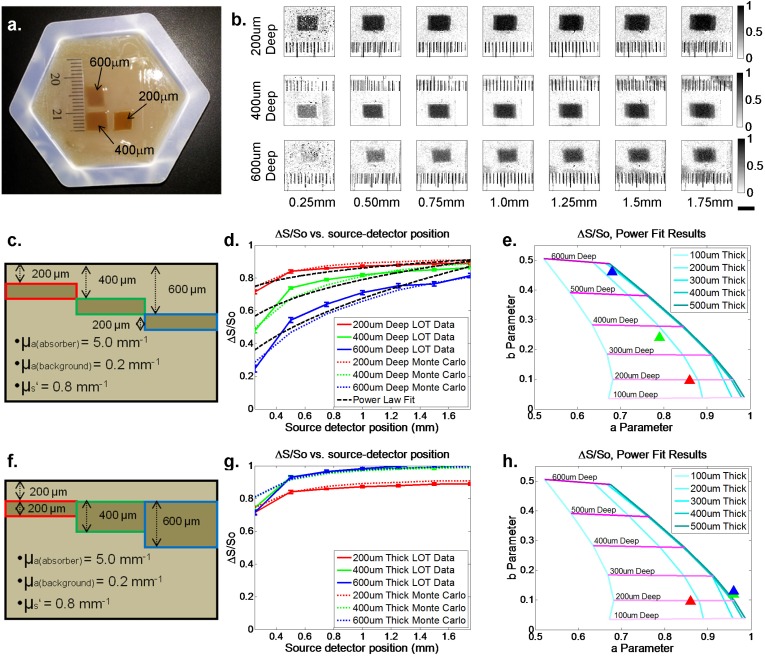
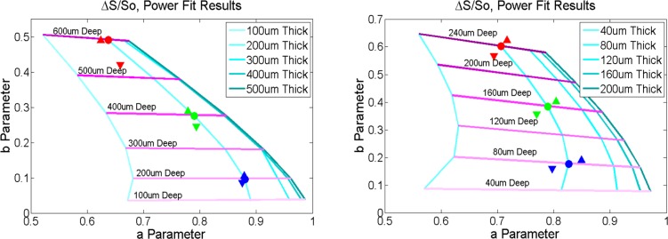
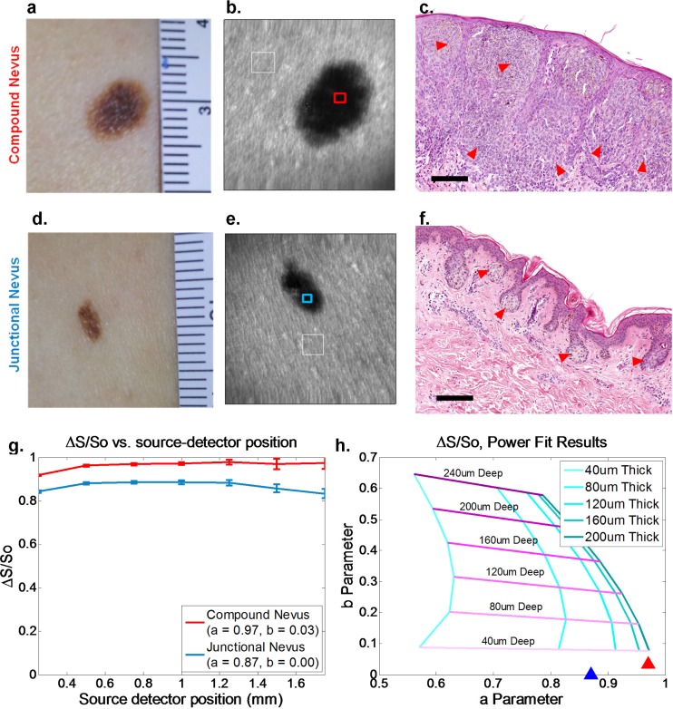
Evaluation of suspicious skin lesions by dermatologists is usually accomplished using white light examination and direct punch or surgical biopsy. However, these techniques can be imprecise for estimating a lesion's margin or level of dermal invasion when planning surgical resection. Laminar optical tomography (LOT) is an imaging technique capable of acquiring depth-sensitive information within scattering tissues. Here, we explore whether LOT data can be used to predict the depth and thickness of pigmented lesions using a range of simulations and phantom models. We then compare these results to LOT data acquired on normal and malignant skin lesions in vivo.
皮肤科医生通常通过白光检查以及直接打孔或手术活检来评估可疑皮肤病变。然而,在规划手术切除时,这些技术在估计病变边缘或真皮浸润程度方面可能并不精确。层流光学断层扫描(LOT)是一种能够在散射组织内获取深度敏感信息的成像技术。在此,我们利用一系列模拟和体模模型探索LOT数据是否可用于预测色素沉着病变的深度和厚度。然后,我们将这些结果与在正常和恶性皮肤病变的体内获取的LOT数据进行比较。