• 文献检索
  • 文档翻译
  • 深度研究
  • 学术资讯
  • Suppr Zotero 插件Zotero 插件
  • 邀请有礼
  • 套餐&价格
  • 历史记录
应用&插件
Suppr Zotero 插件Zotero 插件浏览器插件Mac 客户端Windows 客户端微信小程序
定价
高级版会员购买积分包购买API积分包
服务
文献检索文档翻译深度研究API 文档MCP 服务
关于我们
关于 Suppr公司介绍联系我们用户协议隐私条款
关注我们

Suppr 超能文献

核心技术专利:CN118964589B侵权必究
粤ICP备2023148730 号-1Suppr @ 2026

文献检索

告别复杂PubMed语法,用中文像聊天一样搜索,搜遍4000万医学文献。AI智能推荐,让科研检索更轻松。

立即免费搜索

文件翻译

保留排版,准确专业,支持PDF/Word/PPT等文件格式,支持 12+语言互译。

免费翻译文档

深度研究

AI帮你快速写综述,25分钟生成高质量综述,智能提取关键信息,辅助科研写作。

立即免费体验

骨髓间充质基质细胞通过旁分泌释放 VEGF 刺激骨骼肌成肌细胞增殖。

Bone marrow mesenchymal stromal cells stimulate skeletal myoblast proliferation through the paracrine release of VEGF.

机构信息

Department of Human Anatomy, Histology and Forensic Medicine, University of Florence, Florence, Italy.

出版信息

PLoS One. 2012;7(7):e37512. doi: 10.1371/journal.pone.0037512. Epub 2012 Jul 16.

DOI:10.1371/journal.pone.0037512
PMID:22815682
原文链接:https://pmc.ncbi.nlm.nih.gov/articles/PMC3398011/
Abstract

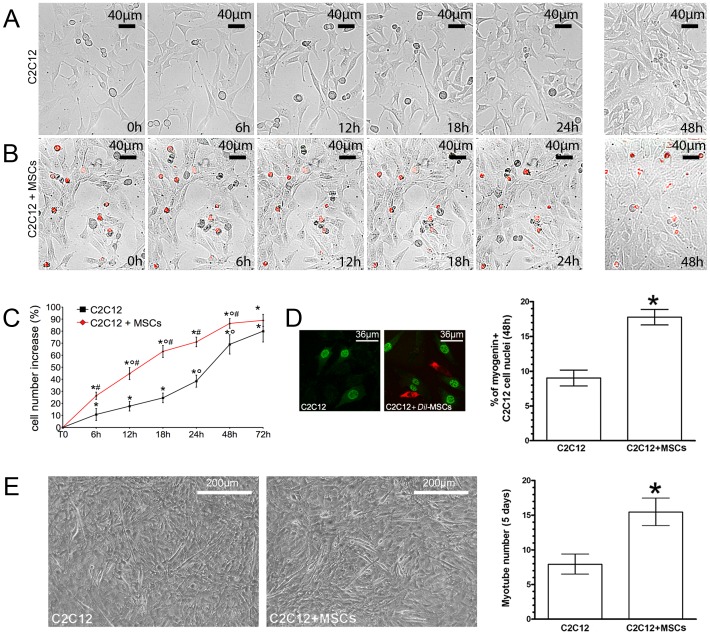
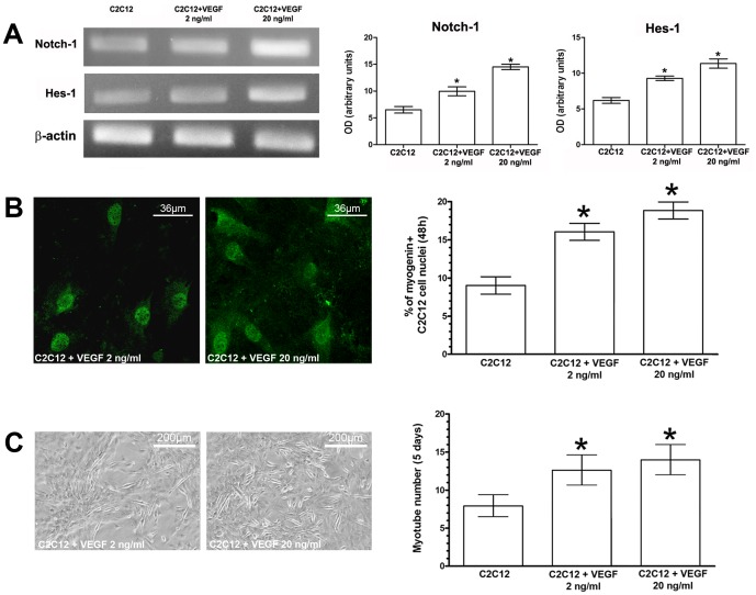
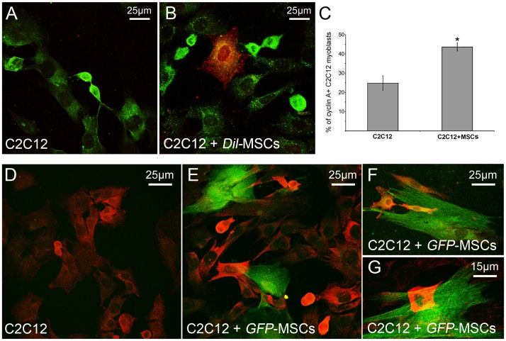
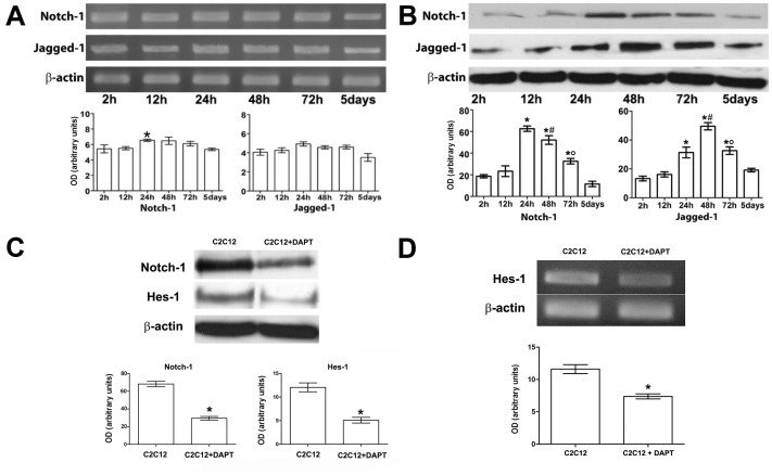
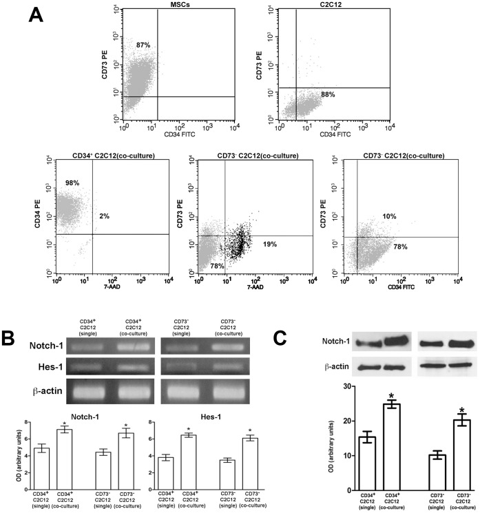
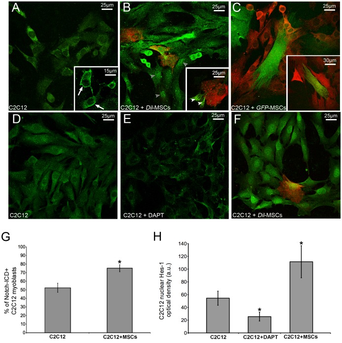
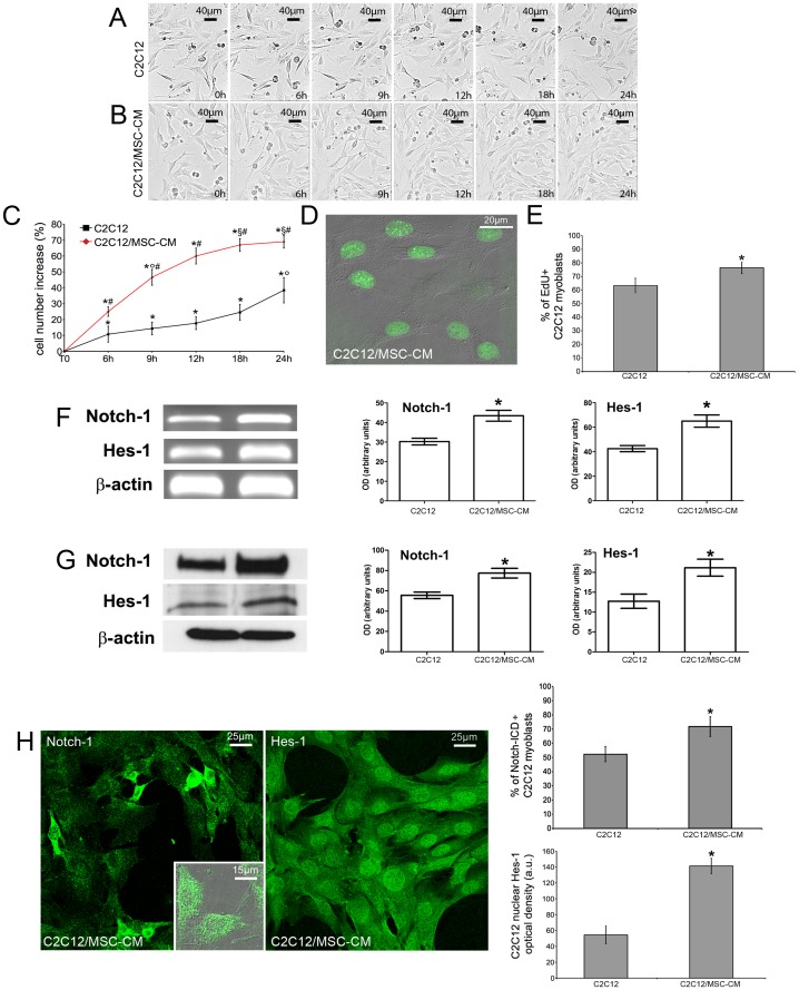
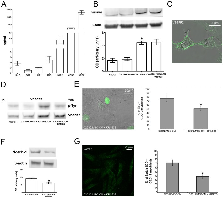
Mesenchymal stromal cells (MSCs) are the leading cell candidates in the field of regenerative medicine. These cells have also been successfully used to improve skeletal muscle repair/regeneration; however, the mechanisms responsible for their beneficial effects remain to be clarified. On this basis, in the present study, we evaluated in a co-culture system, the ability of bone-marrow MSCs to influence C2C12 myoblast behavior and analyzed the cross-talk between the two cell types at the cellular and molecular level. We found that myoblast proliferation was greatly enhanced in the co-culture as judged by time lapse videomicroscopy, cyclin A expression and EdU incorporation. Moreover, myoblasts immunomagnetically separated from MSCs after co-culture expressed higher mRNA and protein levels of Notch-1, a key determinant of myoblast activation and proliferation, as compared with the single culture. Notch-1 intracellular domain and nuclear localization of Hes-1, a Notch-1 target gene, were also increased in the co-culture. Interestingly, the myoblastic response was mainly dependent on the paracrine release of vascular endothelial growth factor (VEGF) by MSCs. Indeed, the addition of MSC-derived conditioned medium (CM) to C2C12 cells yielded similar results as those observed in the co-culture and increased the phosphorylation and expression levels of VEGFR. The treatment with the selective pharmacological VEGFR inhibitor, KRN633, resulted in a marked attenuation of the receptor activation and concomitantly inhibited the effects of MSC-CM on C2C12 cell growth and Notch-1 signaling. In conclusion, this study provides novel evidence for a role of MSCs in stimulating myoblast cell proliferation and suggests that the functional interaction between the two cell types may be exploited for the development of new and more efficient cell-based skeletal muscle repair strategies.

摘要

间充质基质细胞(MSCs)是再生医学领域的主要候选细胞。这些细胞也已成功用于改善骨骼肌修复/再生;然而,负责其有益效果的机制仍需阐明。在此基础上,本研究在共培养体系中评估了骨髓间充质基质细胞影响 C2C12 成肌细胞行为的能力,并在细胞和分子水平上分析了两种细胞类型之间的串扰。我们发现,通过延时视频显微镜、细胞周期蛋白 A 表达和 EdU 掺入判断,共培养中肌母细胞的增殖大大增强。此外,与单一培养相比,从共培养后的 MSC 免疫磁珠分离的肌母细胞表达更高水平的 Notch-1 mRNA 和蛋白,Notch-1 是肌母细胞激活和增殖的关键决定因素。共培养中 Notch-1 细胞内结构域和 Hes-1 的核定位也增加,Hes-1 是 Notch-1 的靶基因。有趣的是,肌母细胞的反应主要依赖于 MSC 旁分泌释放血管内皮生长因子(VEGF)。事实上,将 MSC 衍生的条件培养基(CM)添加到 C2C12 细胞中会产生与共培养中观察到的相似结果,并增加 VEGFR 的磷酸化和表达水平。用选择性 VEGFR 抑制剂 KRN633 处理可显著减弱受体激活,并同时抑制 MSC-CM 对 C2C12 细胞生长和 Notch-1 信号的影响。总之,本研究为 MSC 刺激肌母细胞增殖的作用提供了新的证据,并表明两种细胞类型之间的功能相互作用可能被用于开发新的、更有效的基于细胞的骨骼肌修复策略。

https://cdn.ncbi.nlm.nih.gov/pmc/blobs/6a55/3398011/e9bc63f87ffc/pone.0037512.g008.jpg
https://cdn.ncbi.nlm.nih.gov/pmc/blobs/6a55/3398011/6c7b18132a36/pone.0037512.g001.jpg
https://cdn.ncbi.nlm.nih.gov/pmc/blobs/6a55/3398011/989fbbc91843/pone.0037512.g002.jpg
https://cdn.ncbi.nlm.nih.gov/pmc/blobs/6a55/3398011/0bf5539142ce/pone.0037512.g003.jpg
https://cdn.ncbi.nlm.nih.gov/pmc/blobs/6a55/3398011/9fb44e547904/pone.0037512.g004.jpg
https://cdn.ncbi.nlm.nih.gov/pmc/blobs/6a55/3398011/fc1f50f68f72/pone.0037512.g005.jpg
https://cdn.ncbi.nlm.nih.gov/pmc/blobs/6a55/3398011/d2fb6eb163e0/pone.0037512.g006.jpg
https://cdn.ncbi.nlm.nih.gov/pmc/blobs/6a55/3398011/2b70dbae89eb/pone.0037512.g007.jpg
https://cdn.ncbi.nlm.nih.gov/pmc/blobs/6a55/3398011/e9bc63f87ffc/pone.0037512.g008.jpg

相似文献

1
Bone marrow mesenchymal stromal cells stimulate skeletal myoblast proliferation through the paracrine release of VEGF.骨髓间充质基质细胞通过旁分泌释放 VEGF 刺激骨骼肌成肌细胞增殖。
PLoS One. 2012;7(7):e37512. doi: 10.1371/journal.pone.0037512. Epub 2012 Jul 16.
2
Mesenchymal stromal cell secreted sphingosine 1-phosphate (S1P) exerts a stimulatory effect on skeletal myoblast proliferation.间充质基质细胞分泌的1-磷酸鞘氨醇(S1P)对骨骼肌成肌细胞增殖具有刺激作用。
PLoS One. 2014 Sep 29;9(9):e108662. doi: 10.1371/journal.pone.0108662. eCollection 2014.
3
Combined use of bone marrow-derived mesenchymal stromal cells (BM-MSCs) and platelet rich plasma (PRP) stimulates proliferation and differentiation of myoblasts in vitro: new therapeutic perspectives for skeletal muscle repair/regeneration.骨髓间充质基质细胞(BM-MSCs)和富含血小板的血浆(PRP)的联合使用可刺激体外成肌细胞的增殖和分化:骨骼肌修复/再生的新治疗前景。
Cell Tissue Res. 2018 Jun;372(3):549-570. doi: 10.1007/s00441-018-2792-3. Epub 2018 Feb 5.
4
A Type 2 Deiodinase-Dependent Increase in Mediates Myoblast-Endothelial Cell Crosstalk During Skeletal Muscle Regeneration.一种 2 型脱碘酶依赖性增加的在骨骼肌再生过程中介导成肌细胞-内皮细胞串扰。
Thyroid. 2021 Jan;31(1):115-127. doi: 10.1089/thy.2020.0291. Epub 2020 Sep 9.
5
Mesenchymal Stem Cells Alter the Inflammatory Response of C2C12 Mouse Skeletal Muscle Cells.间充质干细胞改变 C2C12 小鼠骨骼肌细胞的炎症反应。
Biol Pharm Bull. 2020;43(11):1785-1791. doi: 10.1248/bpb.b20-00536.
6
Bone marrow-derived stem cell interactions with adult cardiomyocytes and skeletal myoblasts in vitro.体外骨髓源性干细胞与成年心肌细胞和骨骼肌成肌细胞的相互作用。
Cardiovasc Revasc Med. 2006 Oct-Dec;7(4):222-30. doi: 10.1016/j.carrev.2006.06.005.
7
Mesenchymal stromal cells affect cardiomyocyte growth through juxtacrine Notch-1/Jagged-1 signaling and paracrine mechanisms: clues for cardiac regeneration.间质基质细胞通过旁分泌 Notch-1/Jagged-1 信号和旁分泌机制影响心肌细胞的生长:心脏再生的线索。
J Mol Cell Cardiol. 2011 Sep;51(3):399-408. doi: 10.1016/j.yjmcc.2011.06.004. Epub 2011 Jun 13.
8
The factors present in regenerating muscles impact bone marrow-derived mesenchymal stromal/stem cell fusion with myoblasts.再生肌肉中存在的因素会影响骨髓间充质基质/干细胞与成肌细胞的融合。
Stem Cell Res Ther. 2019 Nov 21;10(1):343. doi: 10.1186/s13287-019-1444-1.
9
Bone marrow-derived mesenchymal stem cells from early diffuse systemic sclerosis exhibit a paracrine machinery and stimulate angiogenesis in vitro.骨髓间充质干细胞从早期弥漫性系统性硬化症表现出一种旁分泌机制,并刺激体外血管生成。
Ann Rheum Dis. 2011 Nov;70(11):2011-21. doi: 10.1136/ard.2011.150607. Epub 2011 Aug 6.
10
[In vitro differentiation of rat mesenchymal stem cells into skeletal muscle cells induced by myoblast differentiation factor and 5-azacytidine].[成肌细胞分化因子和5-氮杂胞苷诱导大鼠间充质干细胞向骨骼肌细胞的体外分化]
Zhongguo Xiu Fu Chong Jian Wai Ke Za Zhi. 2007 Dec;21(12):1371-5.

引用本文的文献

1
Dual Delivery of Cells and Bioactive Molecules for Wound Healing Applications.用于伤口愈合应用的细胞与生物活性分子的双重递送
Molecules. 2025 Mar 31;30(7):1577. doi: 10.3390/molecules30071577.
2
Diversity in Notch ligand-receptor signaling interactions.Notch配体-受体信号相互作用的多样性。
Elife. 2025 Jan 3;12:RP91422. doi: 10.7554/eLife.91422.
3
HUC-MSCs combined with platelet lysate treat diabetic chronic cutaneous ulcers in Bama miniature pig.人脐带间充质干细胞联合血小板裂解物治疗巴马小型猪糖尿病慢性皮肤溃疡

本文引用的文献

1
Therapeutic factors secreted by mesenchymal stromal cells and tissue repair.间充质基质细胞分泌的治疗因子与组织修复。
J Cell Biochem. 2011 Nov;112(11):3073-8. doi: 10.1002/jcb.23250.
2
Mesenchymal stromal cells affect cardiomyocyte growth through juxtacrine Notch-1/Jagged-1 signaling and paracrine mechanisms: clues for cardiac regeneration.间质基质细胞通过旁分泌 Notch-1/Jagged-1 信号和旁分泌机制影响心肌细胞的生长:心脏再生的线索。
J Mol Cell Cardiol. 2011 Sep;51(3):399-408. doi: 10.1016/j.yjmcc.2011.06.004. Epub 2011 Jun 13.
3
The therapeutic potential of bone marrow-derived stromal cells.
Regen Ther. 2024 Nov 20;26:1138-1149. doi: 10.1016/j.reth.2024.11.005. eCollection 2024 Jun.
4
Dietary recommendations of the Moroccan Society of Rheumatology (SMR) for patients with ostéosarcopenia.摩洛哥风湿病学会(SMR)对骨质疏松性肌少症患者的饮食建议。
Arch Osteoporos. 2024 Nov 9;19(1):113. doi: 10.1007/s11657-024-01461-9.
5
Role of mesenchymal stem cells in sepsis and their therapeutic potential in sepsis‑associated myopathy (Review).间充质干细胞在脓毒症中的作用及其在脓毒症相关性肌病中的治疗潜力(综述)。
Int J Mol Med. 2024 Nov;54(5). doi: 10.3892/ijmm.2024.5416. Epub 2024 Sep 2.
6
HIF-1α/MMP-9 Axis Is Required in the Early Phases of Skeletal Myoblast Differentiation under Normoxia Condition In Vitro.在常氧条件下体外骨骼肌成肌细胞分化早期阶段,HIF-1α/MMP-9轴是必需的。
Cells. 2023 Dec 16;12(24):2851. doi: 10.3390/cells12242851.
7
Analysis of the association between osteoporosis and muscle strength in Korean adults: a national cross-sectional study.分析韩国成年人骨质疏松症与肌肉力量之间的关系:一项全国性横断面研究。
J Health Popul Nutr. 2023 Sep 12;42(1):97. doi: 10.1186/s41043-023-00443-w.
8
SDF-1 and NOTCH signaling in myogenic cell differentiation: the role of miRNA10a, 425, and 5100.SDF-1 和 NOTCH 信号在肌源性细胞分化中的作用:miRNA10a、425 和 5100 的作用。
Stem Cell Res Ther. 2023 Aug 15;14(1):204. doi: 10.1186/s13287-023-03429-x.
9
Contribution of peripheral blood mononuclear cells isolated by advanced filtration system to myogenesis of human bone marrow mesenchymal stem cells co-cultured with myoblasts.先进过滤系统分离的外周血单个核细胞对与成肌细胞共培养的人骨髓间充质干细胞成肌作用的贡献。
Heliyon. 2023 Jun 9;9(6):e17141. doi: 10.1016/j.heliyon.2023.e17141. eCollection 2023 Jun.
10
Irisin Is Target of Sphingosine-1-Phosphate/Sphingosine-1-Phosphate Receptor-Mediated Signaling in Skeletal Muscle Cells.鸢尾素是骨骼肌细胞中鞘氨醇-1-磷酸/鞘氨醇-1-磷酸受体介导信号的靶标。
Int J Mol Sci. 2023 Jun 23;24(13):10548. doi: 10.3390/ijms241310548.
骨髓基质细胞的治疗潜力。
J Cell Biochem. 2011 Oct;112(10):2683-7. doi: 10.1002/jcb.23216.
4
Regeneration of skeletal muscle.骨骼肌再生。
Cell Tissue Res. 2012 Mar;347(3):759-74. doi: 10.1007/s00441-011-1185-7. Epub 2011 Jun 11.
5
Overexpression of VEGF165b, an inhibitory splice variant of vascular endothelial growth factor, leads to insufficient angiogenesis in patients with systemic sclerosis.血管内皮生长因子(VEGF)165b 的过表达是一种血管内皮生长因子的抑制性剪接变异体,可导致系统性硬化症患者的血管生成不足。
Circ Res. 2011 Jul 22;109(3):e14-26. doi: 10.1161/CIRCRESAHA.111.242057. Epub 2011 Jun 2.
6
Mesenchymal stem cells in a transgenic mouse model of multiple system atrophy: immunomodulation and neuroprotection.多系统萎缩转基因小鼠模型中的间充质干细胞:免疫调节和神经保护。
PLoS One. 2011;6(5):e19808. doi: 10.1371/journal.pone.0019808. Epub 2011 May 18.
7
Identification of telocytes in skeletal muscle interstitium: implication for muscle regeneration.鉴定骨骼肌间质中的间质细胞:对肌肉再生的影响。
J Cell Mol Med. 2011 Jun;15(6):1379-92. doi: 10.1111/j.1582-4934.2011.01330.x.
8
Mesenchymal stem cells: Molecular characteristics and clinical applications.间质干细胞:分子特征与临床应用。
World J Stem Cells. 2010 Aug 26;2(4):67-80. doi: 10.4252/wjsc.v2.i4.67.
9
Mesenchymal stromal cell therapy: a revolution in Regenerative Medicine?间质基质细胞治疗:再生医学的革命?
Bone Marrow Transplant. 2012 Feb;47(2):164-71. doi: 10.1038/bmt.2011.81. Epub 2011 Apr 11.
10
Effects of S1P on skeletal muscle repair/regeneration during eccentric contraction.S1P 对离心收缩过程中骨骼肌修复/再生的影响。
J Cell Mol Med. 2011 Nov;15(11):2498-511. doi: 10.1111/j.1582-4934.2010.01250.x.