Department of Radiology, University of Tokyo Hospital, 7-3-1 Hongo Bunkyo-ku, Tokyo, Japan.
J Radiat Res. 2012 Jul;53(4):628-32. doi: 10.1093/jrr/rrs013.
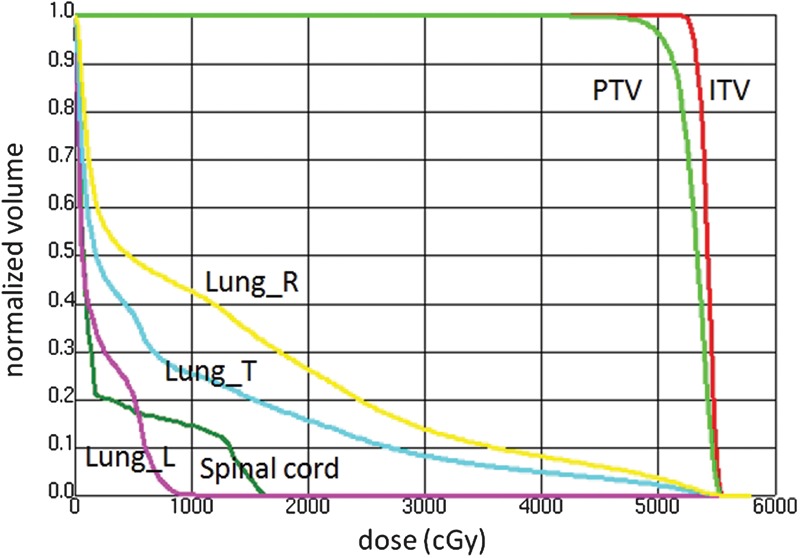
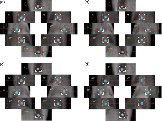
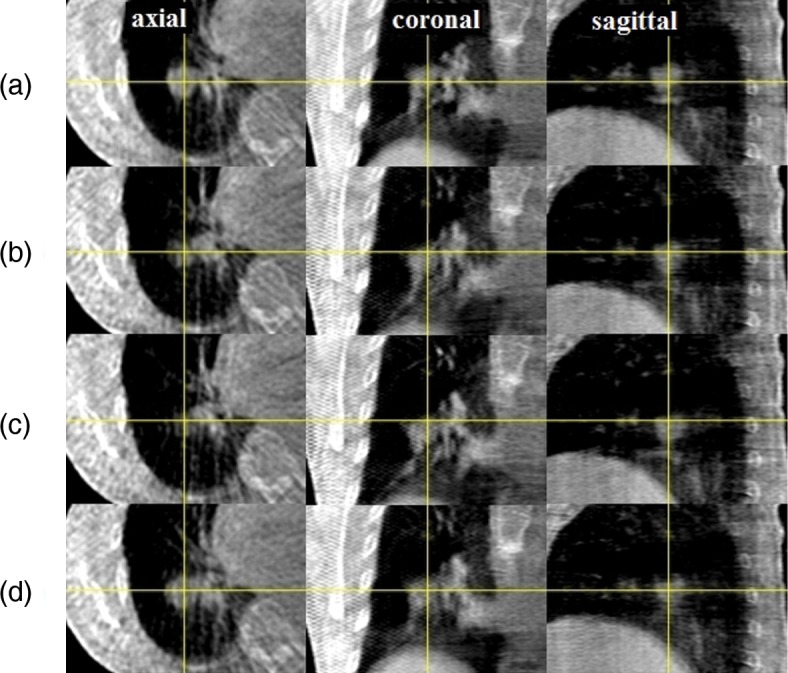
We have proposed four dimensional (4D) digitally reconstructed radiography (DRR) for verifying a lung tumor position during volumetric modulated arc therapy (VMAT). An internal target volume (ITV) was defined based on two clinical target volumes (CTVs) delineated on maximum exhalation and maximum inhalation images acquired by 4D planning computed tomography (CT). A planning target volume (PTV) was defined by adding a margin of 5 mm to the ITV on the maximum exhalation 3D CT images. A single-arc VMAT plan was created on the same CT data using Pinnacle SmartArc with a maximum multi-leaf collimator leaf speed of 1 mm/degree, thereby resulting in quasi-conformal field shapes while optimizing each beam intensity for each gantry angle. During VMAT delivery, cone-beam CT (CBCT) projection data were acquired by an on-board kilovoltage X-ray unit and a flat panel 2D detector. Four CBCT image sets with different respiratory phases were reconstructed using in-house software, where respiratory phases were extracted from the projection data. Subsequently a CTV was delineated on each of the 4D CBCT images by an oncologist. Using the resulting 4D CBCT data including the CTV contours, 4D DRRs during the VMAT delivery were calculated as a function of gantry angle. It was confirmed that the contoured CTV was within the radiation field during the four-fraction lung VMAT delivery. The proposed 4D DRR may facilitate the verification of the position of a respiratory moving lung tumor during VMAT delivery on each treatment day.
我们提出了四维(4D)数字重建放射摄影术(DRR),用于验证容积调强弧形治疗(VMAT)期间肺部肿瘤的位置。基于通过 4D 计划 CT 采集的最大呼气和最大吸气图像上勾画的两个临床靶区(CTV),定义了内部靶区(ITV)。通过在最大呼气 3D CT 图像上向 ITV 添加 5mm 的边界,定义了计划靶区(PTV)。使用 Pinnacle SmartArc 在相同的 CT 数据上创建了单弧 VMAT 计划,最大多叶准直器叶片速度为 1mm/度,从而在优化每个射束强度的同时实现准共形射野形状。在 VMAT 输送过程中,通过机载千伏 X 射线单元和平板二维探测器采集锥形束 CT(CBCT)投影数据。使用内部软件重建了四个具有不同呼吸相位的 CBCT 图像集,其中从投影数据中提取了呼吸相位。随后,由肿瘤学家在每个 4D CBCT 图像上勾画 CTV。使用包括 CTV 轮廓的 4D CBCT 数据,计算了 VMAT 输送过程中的 4D DRR,作为射束角度的函数。证实了在四部分肺部 VMAT 输送过程中,勾画的 CTV 位于辐射场内。所提出的 4D DRR 可能有助于在每个治疗日验证 VMAT 输送过程中呼吸运动肺部肿瘤的位置。