Department of Oncology, University of Calgary, Calgary, Canada.
BMC Cancer. 2012 Aug 1;12:332. doi: 10.1186/1471-2407-12-332.
Resistance to apoptosis is a hallmark of cancer and proteins regulating apoptosis have been proposed as prognostic markers in several malignancies. However, the prognostic impact of apoptotic markers has not been consistently demonstrated in oral squamous cell carcinoma (OSCC). This inconsistency in reported associations between apoptotic proteins and prognosis can be partly attributed to the intrinsic low resolution and misclassification associated with manual, semi-quantitative methods of biomarker expression measurement. The aim of this study was to examine the association between apoptosis-regulating proteins and clinical outcomes in oral squamous cell carcinoma (OSCC) using the quantitative fluorescence immunohistochemistry (IHC) based AQUAnalysis technique.
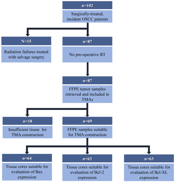
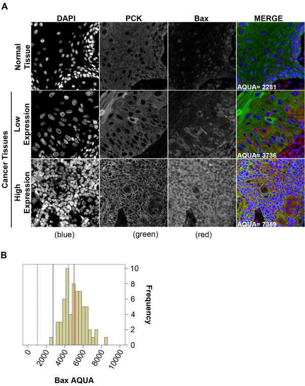
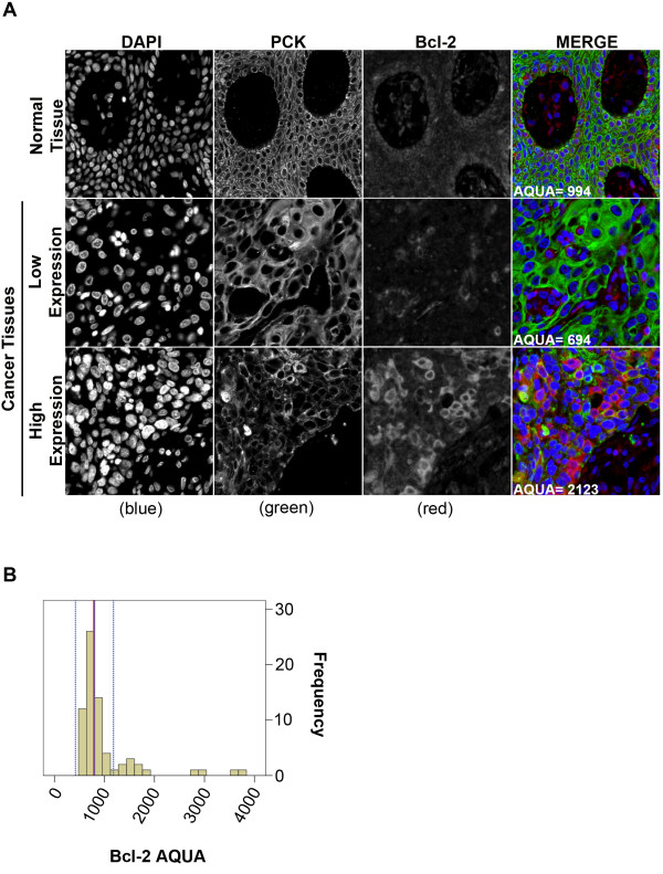
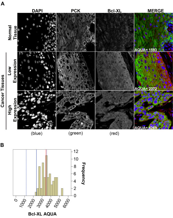
Sixty-nine OSCC patients diagnosed between 1998-2005 in Calgary, Alberta, Canada were included in the study. Clinical data were obtained from the Alberta Cancer Registry and chart review. Tissue microarrays (TMAs) were assembled from triplicate cores of formalin-fixed paraffin embedded pre-treatment tumour tissue. Bax, Bcl-2 and Bcl-XL protein expression was quantified using fluorescent IHC and AQUA technology in normal oral cavity squamous epithelium (OCSE) and OSCC tumour samples. Survival was analyzed using Kaplan-Meier plots and the Cox proportional hazard model.
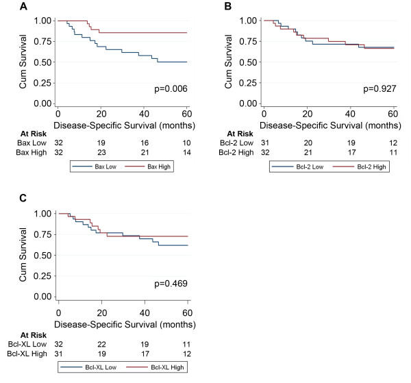
Bax expression was predominantly nuclear in OCSE and almost exclusively cytoplasmic in OSCC. No similar differences in localization were observed for Bcl-2 or Bcl-XL. Only Bax expression associated with disease-specific survival (DSS), with 5-year survival estimates of 85.7% for high Bax versus 50.3% for low Bax (p = 0.006), in univariate analysis. High Bax expression was also significantly associated with elevated Ki67 expression, indicating that increased proliferation might lead to an improved response to radiotherapy in patients with elevated Bax expression. In multivariate analyses, Bax protein expression remained an independent predictor of DSS in OSCC [HR 0.241 (0.078-0.745), p = 0.013].
The AQUA technique used in our study eliminates observer bias and provides reliable and reproducible estimates for biomarker expression. AQUA also provides essential measures of quality control that cannot be achieved with manual biomarker scoring techniques. Our results support the use of Bax protein expression as a prognostic marker in conjunction with other clinico-pathological variables when designing personalized treatment strategies for OSCC patients.
细胞凋亡抵抗是癌症的一个标志,并且已经提出了调节细胞凋亡的蛋白质作为几种恶性肿瘤的预后标志物。然而,凋亡标志物在口腔鳞状细胞癌(OSCC)中的预后影响并不一致。这种报道的凋亡蛋白与预后之间的关联不一致,部分归因于手动、半定量生物标志物表达测量方法固有的低分辨率和分类错误。本研究的目的是使用定量荧光免疫组织化学(IHC)基于 AQUAnalysis 技术检查凋亡调节蛋白与口腔鳞状细胞癌(OSCC)临床结局之间的关联。
本研究纳入了 1998 年至 2005 年在加拿大阿尔伯塔省卡尔加里诊断的 69 例 OSCC 患者。临床数据来自阿尔伯塔癌症登记处和图表审查。组织微阵列(TMA)由福尔马林固定石蜡包埋预处理肿瘤组织的三重复制核心组成。使用荧光 IHC 和 AQUA 技术在正常口腔鳞状上皮(OCSE)和 OSCC 肿瘤样本中定量 Bax、Bcl-2 和 Bcl-XL 蛋白表达。使用 Kaplan-Meier 图和 Cox 比例风险模型分析生存情况。
Bax 表达在 OCSE 中主要为核,而在 OSCC 中几乎完全为细胞质。Bcl-2 或 Bcl-XL 没有观察到类似的定位差异。只有 Bax 表达与疾病特异性生存(DSS)相关,高 Bax 组的 5 年生存率估计为 85.7%,低 Bax 组为 50.3%(p=0.006),在单因素分析中。高 Bax 表达也与 Ki67 表达升高显著相关,表明在高 Bax 表达的患者中,增加增殖可能导致对放疗的反应改善。在多因素分析中,Bax 蛋白表达仍然是 OSCC 中 DSS 的独立预测因子[HR 0.241(0.078-0.745),p=0.013]。
我们研究中使用的 AQUA 技术消除了观察者偏见,并为生物标志物表达提供了可靠和可重复的估计。AQUA 还提供了手动生物标志物评分技术无法实现的重要质量控制措施。我们的结果支持在设计 OSCC 患者的个体化治疗策略时,将 Bax 蛋白表达与其他临床病理变量一起用作预后标志物。