Yokochi Midori, Li Danjie, Horiguchi Masayuki, Kishi Shoji
Department of Ophthalmology, Gunma University School of Medicine, 3-39-22 Showamachi, Maebashi, 371-8511, Japan.
Department of Ophthalmology, Fujita Health University School of Medicine, Aichi, Japan.
Doc Ophthalmol. 2012 Dec;125(3):211-8. doi: 10.1007/s10633-012-9348-8. Epub 2012 Aug 5.
To determine the characteristics of the photoreceptor abnormalities in retinitis pigmentosa (RP) and cone-rod dystrophy (CRD).
We evaluated the photoreceptor abnormalities using spectral-domain optical coherence tomography (SD-OCT) in 28 patients with RP and 17 patients with CRD. The OCT images and full-field electroretinograms were obtained from 21 eyes in normal subjects who were age-matched to patients with RP and CRD and served as controls.
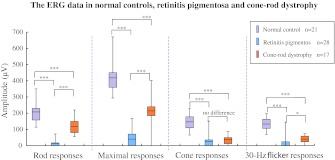
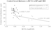
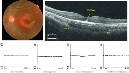
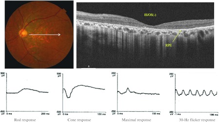
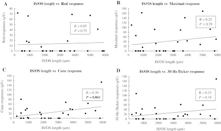
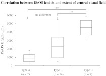
Eyes with RP and CRD had markedly decreased rod responses (6.5 and 57.5 % of normal value), maximal responses (9.6 and 51.6 %), cone (16.5 and 25.8 %), and 30-Hz flicker responses (17.8 and 30.1 % of normal value), and their P values were smaller than 0.0003. On comparison of ERG data between RP and CRD, they had statistically significant differences in rod responses (P < 0.0003) and maximal responses (P < 0.0003). However, there were no statistical differences in cone response and a weak difference in 30-Hz flicker responses (P < 0.017). The best-corrected visual acuity was -0.03 ± 0.09 (logMAR, mean ± standard deviation [SD]) in eyes with RP, but 0.57 ± 0.54 in eyes with CRD. SD-OCT showed that eyes with RP had an intact reflective line at the junction between the photoreceptor inner and outer segment (IS/OS) at the fovea, while eyes with CRD had no IS/OS. The extent of the central visual field was correlated with the IS/OS length at the macula in eyes with RP.
The distribution patterns of the IS/OS line help to differentiate between RP and CRD.
确定视网膜色素变性(RP)和锥杆营养不良(CRD)中光感受器异常的特征。
我们使用光谱域光学相干断层扫描(SD-OCT)评估了28例RP患者和17例CRD患者的光感受器异常情况。从年龄与RP和CRD患者相匹配的21名正常受试者的眼睛中获取了OCT图像和全视野视网膜电图,并将其作为对照。
RP和CRD患者的眼睛杆反应(分别为正常值的6.5%和57.5%)、最大反应(9.6%和51.6%)、锥体反应(16.5%和25.8%)以及30赫兹闪烁反应(分别为正常值的17.8%和30.1%)均显著降低,其P值均小于0.0003。比较RP和CRD之间的视网膜电图数据,它们在杆反应(P < 0.0003)和最大反应(P < 0.0003)方面存在统计学显著差异。然而,锥体反应无统计学差异,30赫兹闪烁反应存在微弱差异(P < 0.017)。RP患者眼睛的最佳矫正视力为-0.03 ± 0.09(logMAR,平均值±标准差[SD]),而CRD患者眼睛的最佳矫正视力为0.57 ± 0.54。SD-OCT显示,RP患者的眼睛在黄斑中心凹处光感受器内段和外段(IS/OS)交界处的反射线完整,而CRD患者的眼睛没有IS/OS。RP患者眼睛的中心视野范围与黄斑处的IS/OS长度相关。
IS/OS线的分布模式有助于区分RP和CRD。