Shin Joong Won, Shin Yong Un, Cho Hee Yoon, Lee Byung Ro
Department of Ophthalmology, Hanyang University College of Medicine, Seoul, Korea.
Korean J Ophthalmol. 2012 Aug;26(4):255-9. doi: 10.3341/kjo.2012.26.4.255. Epub 2012 Jul 24.
To study choroidal thickness and its topographic profile in normal eyes using 3D OCT-1000 spectral domain optical coherence tomography and the correlation with age and refractive error.
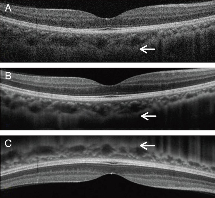
Fifty-seven eyes (45 individuals) with no visual complaints or ocular disease underwent horizontal and vertical line scanning using 3D OCT-1000. The definition of choroidal thickness was the vertical distance between the posterior edge of the hyper-reflective retinal pigment epithelium and the choroid/sclera junction. Choroidal thickness was measured in the subfoveal area at 500 µm intervals from the fovea to 2,500 µm in the nasal, temporal, superior, and inferior regions. The spherical equivalent refractive error was measured by autorefractometry. Statistical analysis was used to confirm the correlations of choroidal thickness with age and refraction error.
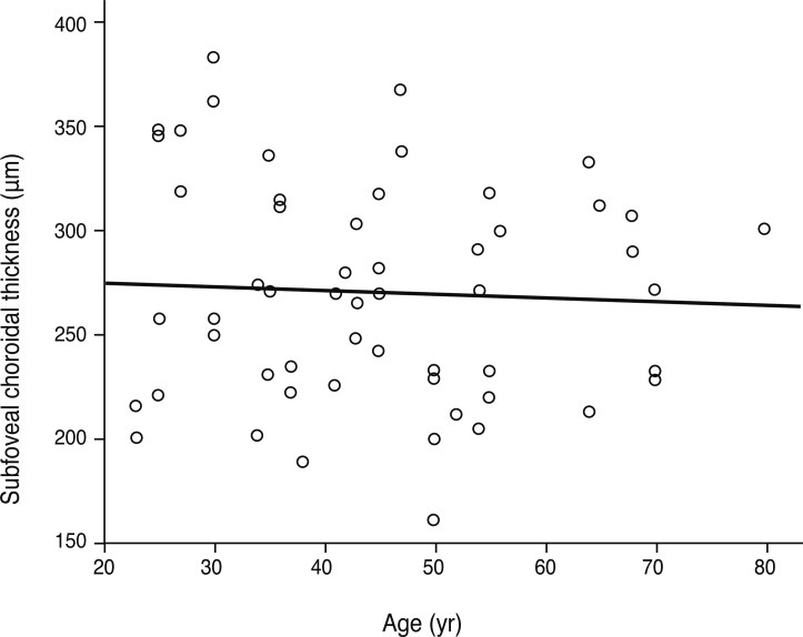
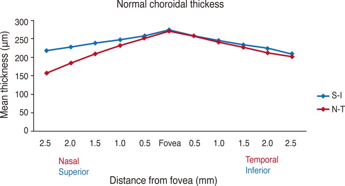
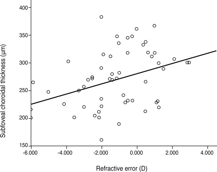
The mean age of the 45 participants (57 eyes) was 45.28 years. Detailed visualization of the choroid for measuring its thickness was possible in 63.3% of eyes. The mean subfoveal choroidal thickness was found to be 270.8 µm (standard deviation [SD], ±51 µm), in horizontal scanning and 275.0 µm (SD, ±49 µm) in vertical scanning. The temporal choroidal thickness was greater than any 500 µm interval in corresponding locations, and there was no significant difference between the superior and inferior choroid as far as 2,000 µm from the fovea. Age and refractive error were associated with subfoveal choroidal thickness in terms of regression (p < 0.05).
Choroidal thickness in normal Korean eyes can be measured using 3D OCT-1000 with high resolution line scanning. The topographical profile of choroidal thickness varies depending on its location. Age and refractive error are essential factors for interpretation of choroidal thickness.
使用3D OCT - 1000光谱域光学相干断层扫描技术研究正常眼脉络膜厚度及其地形图特征,并探讨其与年龄和屈光不正的相关性。
对57只眼(45名个体)进行检查,这些受试者无视觉主诉或眼部疾病,使用3D OCT - 1000进行水平和垂直线扫描。脉络膜厚度定义为高反射性视网膜色素上皮后缘与脉络膜/巩膜交界处之间的垂直距离。在黄斑中心凹下区域,从黄斑中心凹开始,以500 µm的间隔,在鼻侧、颞侧、上方和下方区域测量至2500 µm处的脉络膜厚度。通过自动验光仪测量等效球镜屈光不正。采用统计学分析来确定脉络膜厚度与年龄和屈光不正之间的相关性。
45名参与者(57只眼)的平均年龄为45.28岁。63.3%的眼睛能够清晰显示脉络膜以测量其厚度。水平扫描时黄斑中心凹下脉络膜平均厚度为270.8 µm(标准差[SD],±51 µm),垂直扫描时为275.0 µm(SD,±49 µm)。颞侧脉络膜厚度在相应位置大于任何500 µm间隔处,在距黄斑中心凹2000 µm范围内,上方和下方脉络膜厚度无显著差异。年龄和屈光不正与黄斑中心凹下脉络膜厚度在回归分析方面相关(p < 0.05)。
使用3D OCT - 1000进行高分辨率线扫描可测量正常韩国人眼睛的脉络膜厚度。脉络膜厚度的地形图特征因位置而异。年龄和屈光不正是解释脉络膜厚度的重要因素。