• 文献检索
  • 文档翻译
  • 深度研究
  • 学术资讯
  • Suppr Zotero 插件Zotero 插件
  • 邀请有礼
  • 套餐&价格
  • 历史记录
应用&插件
Suppr Zotero 插件Zotero 插件浏览器插件Mac 客户端Windows 客户端微信小程序
定价
高级版会员购买积分包购买API积分包
服务
文献检索文档翻译深度研究API 文档MCP 服务
关于我们
关于 Suppr公司介绍联系我们用户协议隐私条款
关注我们

Suppr 超能文献

核心技术专利:CN118964589B侵权必究
粤ICP备2023148730 号-1Suppr @ 2026

文献检索

告别复杂PubMed语法,用中文像聊天一样搜索,搜遍4000万医学文献。AI智能推荐,让科研检索更轻松。

立即免费搜索

文件翻译

保留排版,准确专业,支持PDF/Word/PPT等文件格式,支持 12+语言互译。

免费翻译文档

深度研究

AI帮你快速写综述,25分钟生成高质量综述,智能提取关键信息,辅助科研写作。

立即免费体验

光致变色荧光蛋白的焦点切换实现了具有卓越图像对比度的多光子显微镜技术。

Focal switching of photochromic fluorescent proteins enables multiphoton microscopy with superior image contrast.

作者信息

Kao Ya-Ting, Zhu Xinxin, Xu Fang, Min Wei

机构信息

Department of Chemistry, Columbia University, New York, NY 10027, USA.

出版信息

Biomed Opt Express. 2012 Aug 1;3(8):1955-63. doi: 10.1364/BOE.3.001955. Epub 2012 Jul 27.

DOI:10.1364/BOE.3.001955
PMID:22876358
原文链接:https://pmc.ncbi.nlm.nih.gov/articles/PMC3409713/
Abstract

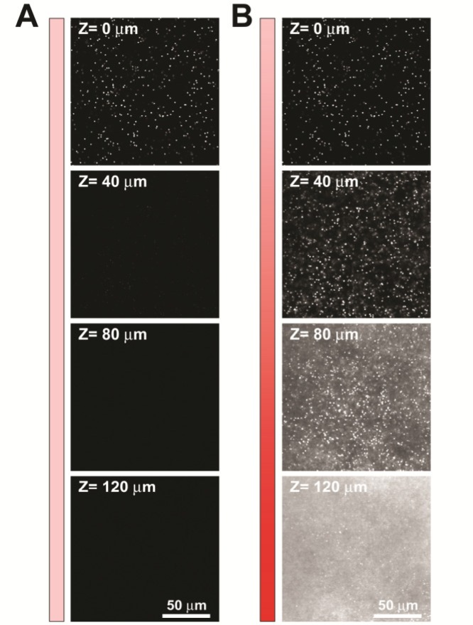
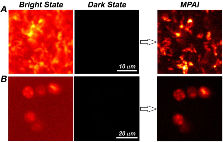
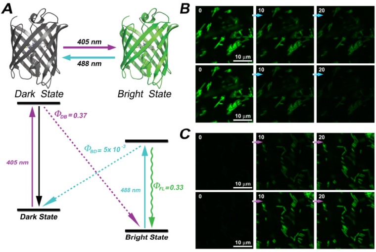
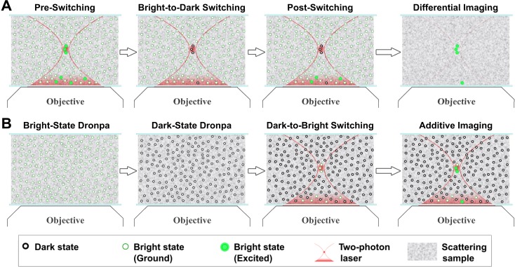
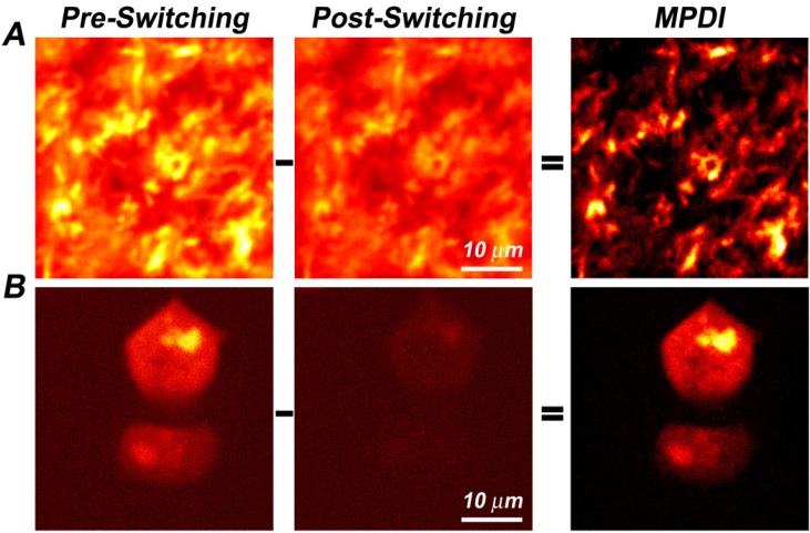
Probing biological structures and functions deep inside live organisms with light is highly desirable. Among the current optical imaging modalities, multiphoton fluorescence microscopy exhibits the best contrast for imaging scattering samples by employing a spatially confined nonlinear excitation. However, as the incident laser power drops exponentially with imaging depth into the sample due to the scattering loss, the out-of-focus background eventually overwhelms the in-focus signal, which defines a fundamental imaging-depth limit. Herein we significantly improve the image contrast for deep scattering samples by harnessing reversibly switchable fluorescent proteins (RSFPs) which can be cycled between bright and dark states upon light illumination. Two distinct techniques, multiphoton deactivation and imaging (MPDI) and multiphoton activation and imaging (MPAI), are demonstrated on tissue phantoms labeled with Dronpa protein. Such a focal switch approach can generate pseudo background-free images. Conceptually different from wave-based approaches that try to reduce light scattering in turbid samples, our work represents a molecule-based strategy that focused on imaging probes.

摘要

利用光深入探测活生物体内部的生物结构和功能是非常有必要的。在当前的光学成像方式中,多光子荧光显微镜通过采用空间受限的非线性激发,在对散射样本成像时展现出最佳的对比度。然而,由于散射损耗,入射激光功率随成像深度呈指数下降,离焦背景最终会淹没聚焦信号,这就定义了一个基本的成像深度极限。在此,我们通过利用可逆开关荧光蛋白(RSFP)显著提高了对深度散射样本的图像对比度,该蛋白在光照下可在明亮和黑暗状态之间循环。在标记有Dronpa蛋白的组织模型上展示了两种不同的技术,即多光子失活与成像(MPDI)和多光子激活与成像(MPAI)。这种焦点切换方法可以生成伪无背景图像。与试图减少浑浊样本中光散射的基于波的方法在概念上不同,我们的工作代表了一种基于分子的策略,专注于成像探针。

https://cdn.ncbi.nlm.nih.gov/pmc/blobs/3f5a/3409713/7ff49014b848/boe-3-8-1955-g005.jpg
https://cdn.ncbi.nlm.nih.gov/pmc/blobs/3f5a/3409713/f37da5f02db6/boe-3-8-1955-g001.jpg
https://cdn.ncbi.nlm.nih.gov/pmc/blobs/3f5a/3409713/8e4e321ae6e5/boe-3-8-1955-g002.jpg
https://cdn.ncbi.nlm.nih.gov/pmc/blobs/3f5a/3409713/3bc2fac1788f/boe-3-8-1955-g003.jpg
https://cdn.ncbi.nlm.nih.gov/pmc/blobs/3f5a/3409713/2c88c46f3540/boe-3-8-1955-g004.jpg
https://cdn.ncbi.nlm.nih.gov/pmc/blobs/3f5a/3409713/7ff49014b848/boe-3-8-1955-g005.jpg

相似文献

1
Focal switching of photochromic fluorescent proteins enables multiphoton microscopy with superior image contrast.光致变色荧光蛋白的焦点切换实现了具有卓越图像对比度的多光子显微镜技术。
Biomed Opt Express. 2012 Aug 1;3(8):1955-63. doi: 10.1364/BOE.3.001955. Epub 2012 Jul 27.
2
Extending the fundamental imaging-depth limit of multi-photon microscopy by imaging with photo-activatable fluorophores.通过使用光激活荧光团成像来扩展多光子显微镜的基本成像深度极限。
Opt Express. 2012 Aug 13;20(17):18525-36. doi: 10.1364/OE.20.018525.
3
Stimulated emission reduced fluorescence microscopy: a concept for extending the fundamental depth limit of two-photon fluorescence imaging.受激发射减少荧光显微镜:一种扩展双光子荧光成像基本深度极限的概念。
Biomed Opt Express. 2012 Jun 1;3(6):1465-75. doi: 10.1364/BOE.3.001465. Epub 2012 May 22.
4
Highly photostable, reversibly photoswitchable fluorescent protein with high contrast ratio for live-cell superresolution microscopy.用于活细胞超分辨率显微镜的具有高光稳定性、可逆光开关特性且对比度高的荧光蛋白。
Proc Natl Acad Sci U S A. 2016 Sep 13;113(37):10364-9. doi: 10.1073/pnas.1611038113. Epub 2016 Aug 25.
5
Imaging deeper than the transport mean free path with multiphoton microscopy.利用多光子显微镜进行比输运平均自由程更深的成像。
Biomed Opt Express. 2021 Dec 21;13(1):452-463. doi: 10.1364/BOE.444696. eCollection 2022 Jan 1.
6
Multiphoton excitation provides optical sections from deeper within scattering specimens than confocal imaging.与共聚焦成像相比,多光子激发能够从散射样本更深的内部提供光学切片。
Biophys J. 1998 Oct;75(4):2015-24. doi: 10.1016/S0006-3495(98)77643-X.
7
Reversibly switchable fluorescence microscopy with enhanced resolution and image contrast.具有更高分辨率和图像对比度的可逆切换荧光显微镜。
J Biomed Opt. 2014 Aug;19(8):086018. doi: 10.1117/1.JBO.19.8.086018.
8
The Positive Switching Fluorescent Protein Padron2 Enables Live-Cell Reversible Saturable Optical Linear Fluorescence Transitions (RESOLFT) Nanoscopy without Sequential Illumination Steps.阳性转换荧光蛋白 Padron2 可实现无需连续激发步骤的活细胞可逆饱和光线性荧光跃迁(RESOLFT)纳米显微镜。
ACS Nano. 2021 Jun 22;15(6):9509-9521. doi: 10.1021/acsnano.0c08207. Epub 2021 May 21.
9
Multiphoton excitation spectra in biological samples.生物样品中的多光子激发光谱。
J Biomed Opt. 2003 Jul;8(3):329-38. doi: 10.1117/1.1583734.
10
Hyperspectral multiphoton microscopy for visualization of multiple, spectrally overlapped fluorescent labels.用于可视化多种光谱重叠荧光标记的高光谱多光子显微镜。
Optica. 2020 Nov 20;7(11):1587-1601. doi: 10.1364/optica.389982.

引用本文的文献

1
Genetically encoded photo-switchable molecular sensors for optoacoustic and super-resolution imaging.基因编码光致开关分子传感器用于光声和超分辨率成像。
Nat Biotechnol. 2022 Apr;40(4):598-605. doi: 10.1038/s41587-021-01100-5. Epub 2021 Nov 29.
2
Two-photon probes for in vivo multicolor microscopy of the structure and signals of brain cells.用于活体内脑细胞结构和信号的双色荧光探针
Brain Struct Funct. 2018 Sep;223(7):3011-3043. doi: 10.1007/s00429-018-1678-1. Epub 2018 May 11.
3
Two-photon-like microscopy with orders-of-magnitude lower illumination intensity via two-step fluorescence.

本文引用的文献

1
Stimulated emission reduced fluorescence microscopy: a concept for extending the fundamental depth limit of two-photon fluorescence imaging.受激发射减少荧光显微镜:一种扩展双光子荧光成像基本深度极限的概念。
Biomed Opt Express. 2012 Jun 1;3(6):1465-75. doi: 10.1364/BOE.3.001465. Epub 2012 May 22.
2
Protein-flexibility mediated coupling between photoswitching kinetics and surrounding viscosity of a photochromic fluorescent protein.蛋白柔性介导的光致变色荧光蛋白的光开关动力学与周围黏度的偶联关系。
Proc Natl Acad Sci U S A. 2012 Feb 28;109(9):3220-5. doi: 10.1073/pnas.1115311109. Epub 2012 Feb 10.
3
Nonlinear structured-illumination microscopy with a photoswitchable protein reveals cellular structures at 50-nm resolution.
通过两步荧光实现照明强度降低几个数量级的类双光子显微镜。
Nat Commun. 2015 Sep 3;6:8184. doi: 10.1038/ncomms9184.
4
A fast- and positively photoswitchable fluorescent protein for ultralow-laser-power RESOLFT nanoscopy.一种快速且正向光开关的荧光蛋白,用于超低激光功率 RESOLFT 纳米显微镜。
Nat Methods. 2015 Jun;12(6):515-8. doi: 10.1038/nmeth.3362. Epub 2015 Apr 20.
5
Reversibly switchable fluorescence microscopy with enhanced resolution and image contrast.具有更高分辨率和图像对比度的可逆切换荧光显微镜。
J Biomed Opt. 2014 Aug;19(8):086018. doi: 10.1117/1.JBO.19.8.086018.
基于光激活蛋白的非线性结构光照明显微镜可实现 50nm 分辨率的细胞结构成像。
Proc Natl Acad Sci U S A. 2012 Jan 17;109(3):E135-43. doi: 10.1073/pnas.1107547108. Epub 2011 Dec 12.
4
In vivo two-photon microscopy to 1.6-mm depth in mouse cortex.活体小鼠皮层内 1.6 毫米深度的双光子显微镜成像。
J Biomed Opt. 2011 Oct;16(10):106014. doi: 10.1117/1.3646209.
5
Diffraction-unlimited all-optical imaging and writing with a photochromic GFP.利用光致变色 GFP 实现无衍射极限的全光学成象和写入。
Nature. 2011 Sep 11;478(7368):204-8. doi: 10.1038/nature10497.
6
Scale: a chemical approach for fluorescence imaging and reconstruction of transparent mouse brain.比例尺:一种用于荧光成像和透明小鼠大脑重建的化学方法。
Nat Neurosci. 2011 Aug 30;14(11):1481-8. doi: 10.1038/nn.2928.
7
Two-photon absorption properties of fluorescent proteins.荧光蛋白的双光子吸收特性。
Nat Methods. 2011 May;8(5):393-9. doi: 10.1038/nmeth.1596. Epub 2011 Apr 28.
8
Maximum imaging depth of two-photon autofluorescence microscopy in epithelial tissues.上皮组织中二光子自发荧光显微镜的最大成像深度。
J Biomed Opt. 2011 Feb;16(2):026008. doi: 10.1117/1.3548646.
9
High-resolution fluorescence microscopy based on a cyclic sequential multiphoton process.基于循环顺序多光子过程的高分辨率荧光显微镜。
Biomed Opt Express. 2010 Sep 7;1(3):791-797. doi: 10.1364/BOE.1.000791.
10
Modern fluorescent proteins and imaging technologies to study gene expression, nuclear localization, and dynamics.现代荧光蛋白和成像技术可用于研究基因表达、核定位和动态变化。
Curr Opin Cell Biol. 2011 Jun;23(3):310-7. doi: 10.1016/j.ceb.2010.12.004. Epub 2011 Jan 15.