• 文献检索
  • 文档翻译
  • 深度研究
  • 学术资讯
  • Suppr Zotero 插件Zotero 插件
  • 邀请有礼
  • 套餐&价格
  • 历史记录
应用&插件
Suppr Zotero 插件Zotero 插件浏览器插件Mac 客户端Windows 客户端微信小程序
定价
高级版会员购买积分包购买API积分包
服务
文献检索文档翻译深度研究API 文档MCP 服务
关于我们
关于 Suppr公司介绍联系我们用户协议隐私条款
关注我们

Suppr 超能文献

核心技术专利:CN118964589B侵权必究
粤ICP备2023148730 号-1Suppr @ 2026

文献检索

告别复杂PubMed语法,用中文像聊天一样搜索,搜遍4000万医学文献。AI智能推荐,让科研检索更轻松。

立即免费搜索

文件翻译

保留排版,准确专业,支持PDF/Word/PPT等文件格式,支持 12+语言互译。

免费翻译文档

深度研究

AI帮你快速写综述,25分钟生成高质量综述,智能提取关键信息,辅助科研写作。

立即免费体验

基于超高时间分辨率相位对比心血管磁共振的波强分析的无创临床应用。

A non-invasive clinical application of wave intensity analysis based on ultrahigh temporal resolution phase-contrast cardiovascular magnetic resonance.

机构信息

Centre for Cardiovascular Imaging, UCL Institute of Cardiovascular Science, and Great Ormond Street Hospital for Children, NHS Trust, London, UK.

出版信息

J Cardiovasc Magn Reson. 2012 Aug 9;14(1):57. doi: 10.1186/1532-429X-14-57.

DOI:10.1186/1532-429X-14-57
PMID:22876747
原文链接:https://pmc.ncbi.nlm.nih.gov/articles/PMC3472227/
Abstract

BACKGROUND

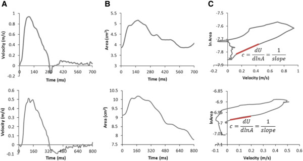
Wave intensity analysis, traditionally derived from pressure and velocity data, can be formulated using velocity and area. Flow-velocity and area can both be derived from high-resolution phase-contrast cardiovascular magnetic resonance (PC-CMR). In this study, very high temporal resolution PC-CMR data is processed using an integrated and semi-automatic technique to derive wave intensity.

METHODS

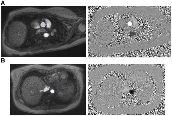
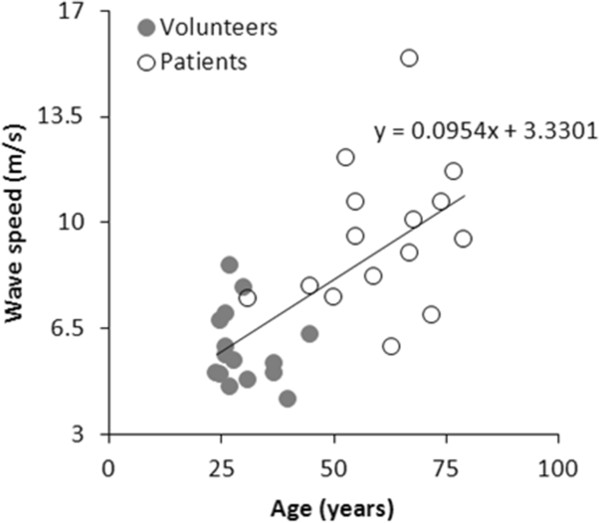
Wave intensity was derived in terms of area and velocity changes. These data were directly derived from PC-CMR using a breath-hold spiral sequence accelerated with sensitivity encoding (SENSE). Image processing was integrated in a plug-in for the DICOM viewer OsiriX, including calculations of wave speed and wave intensity. Ascending and descending aortic data from 15 healthy volunteers (30 ± 6 years) data were used to test the method for feasibility, and intra- and inter-observer variability. Ascending aortic data were also compared with results from 15 patients with coronary heart disease (61 ± 13 years) to assess the clinical usefulness of the method.

RESULTS

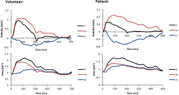
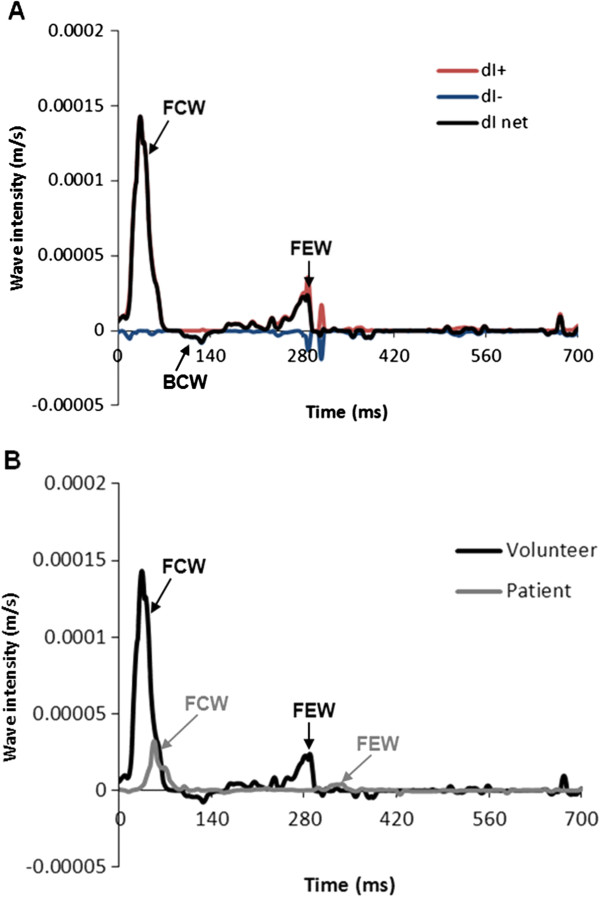
Rapid image acquisition (11 s breath-hold) and image processing was feasible in all volunteers. Wave speed was physiological (5.8 ± 1.3 m/s ascending aorta, 5.0 ± 0.7 m/s descending aorta) and the wave intensity pattern was consistent with traditionally formulated wave intensity. Wave speed, peak forward compression wave in early systole and peak forward expansion wave in late systole at both locations exhibited overall good intra- and inter-observer variability. Patients with coronary heart disease had higher wave speed (p <0.0001), and lower forward compression wave (p <0.0001) and forward expansion wave (p <0.0005) peaks. This difference is likely related to the older age of the patients' cohort, indicating stiffer aortas, as well as compromised ventricular function due to their underlying condition.

CONCLUSION

A non-invasive, semi-automated and reproducible method for performing wave intensity analysis is presented. Its application is facilitated by the use of a very high temporal resolution spiral sequence. A formulation of wave intensity based on area change has also been proposed, involving no assumptions about the cross-sectional shape of the vessel.

摘要

背景

传统的波强分析是基于压力和速度数据推导出来的,也可以基于速度和面积来构建。流速和面积都可以从高分辨率相位对比心血管磁共振(PC-CMR)中得到。在本研究中,使用集成和半自动技术处理超高时间分辨率 PC-CMR 数据,以推导波强。

方法

波强是根据面积和速度变化推导出来的。这些数据是直接从使用呼吸门控螺旋序列加速灵敏度编码(SENSE)的 PC-CMR 中获得的。图像处理集成在 DICOM 查看器 OsiriX 的插件中,包括波速和波强的计算。15 名健康志愿者(30±6 岁)的升主动脉和降主动脉数据用于测试该方法的可行性和观察者内及观察者间的可变性。还将升主动脉数据与 15 名冠心病患者(61±13 岁)的结果进行比较,以评估该方法的临床应用价值。

结果

所有志愿者都可以快速采集图像(11s 屏气)和进行图像处理。波速是生理性的(升主动脉 5.8±1.3m/s,降主动脉 5.0±0.7m/s),波强模式与传统的波强一致。两个部位的波速、早期收缩期正向压缩波峰值和晚期收缩期正向扩张波峰值都具有较好的观察者内和观察者间的可变性。冠心病患者的波速较高(p<0.0001),正向压缩波峰值和正向扩张波峰值较低(p<0.0001 和 p<0.0005)。这种差异可能与患者年龄较大有关,表明主动脉僵硬,以及由于基础疾病导致心室功能受损。

结论

本文提出了一种非侵入性、半自动和可重复的波强分析方法。该方法通过使用超高时间分辨率的螺旋序列来简化其应用。还提出了一种基于面积变化的波强公式,该公式不涉及对血管横截面形状的任何假设。

https://cdn.ncbi.nlm.nih.gov/pmc/blobs/d31c/3472227/954359637d87/1532-429X-14-57-5.jpg
https://cdn.ncbi.nlm.nih.gov/pmc/blobs/d31c/3472227/08d14e981fe7/1532-429X-14-57-1.jpg
https://cdn.ncbi.nlm.nih.gov/pmc/blobs/d31c/3472227/9de5794fc63a/1532-429X-14-57-2.jpg
https://cdn.ncbi.nlm.nih.gov/pmc/blobs/d31c/3472227/0bebba22b954/1532-429X-14-57-3.jpg
https://cdn.ncbi.nlm.nih.gov/pmc/blobs/d31c/3472227/108ebc1e0f0b/1532-429X-14-57-4.jpg
https://cdn.ncbi.nlm.nih.gov/pmc/blobs/d31c/3472227/954359637d87/1532-429X-14-57-5.jpg

相似文献

1
A non-invasive clinical application of wave intensity analysis based on ultrahigh temporal resolution phase-contrast cardiovascular magnetic resonance.基于超高时间分辨率相位对比心血管磁共振的波强分析的无创临床应用。
J Cardiovasc Magn Reson. 2012 Aug 9;14(1):57. doi: 10.1186/1532-429X-14-57.
2
Feasibility of cardiovascular magnetic resonance derived coronary wave intensity analysis.心血管磁共振衍生的冠状动脉波强度分析的可行性
J Cardiovasc Magn Reson. 2016 Dec 9;18(1):93. doi: 10.1186/s12968-016-0312-8.
3
Comparison of different methods for the estimation of aortic pulse wave velocity from 4D flow cardiovascular magnetic resonance.从 4D 流心血管磁共振评估主动脉脉搏波速度的不同方法比较。
J Cardiovasc Magn Reson. 2019 Dec 12;21(1):75. doi: 10.1186/s12968-019-0584-x.
4
Aortic length measurements for pulse wave velocity calculation: manual 2D vs automated 3D centreline extraction.用于脉搏波速度计算的主动脉长度测量:手动二维与自动三维中心线提取
J Cardiovasc Magn Reson. 2017 Mar 8;19(1):32. doi: 10.1186/s12968-017-0341-y.
5
Regional assessment of carotid artery pulse wave velocity using compressed sensing accelerated high temporal resolution 2D CINE phase contrast cardiovascular magnetic resonance.应用压缩感知加速高时间分辨率二维 Cine 相位对比心血管磁共振技术对颈动脉脉搏波速度进行区域性评估。
J Cardiovasc Magn Reson. 2018 Dec 20;20(1):86. doi: 10.1186/s12968-018-0499-y.
6
Required temporal resolution for accurate thoracic aortic pulse wave velocity measurements by phase-contrast magnetic resonance imaging and comparison with clinical standard applanation tonometry.通过相位对比磁共振成像准确测量胸主动脉脉搏波速度所需的时间分辨率以及与临床标准压平式眼压计的比较。
BMC Cardiovasc Disord. 2016 May 26;16(1):110. doi: 10.1186/s12872-016-0292-5.
7
Assessment of local pulse wave velocity distribution in mice using k-t BLAST PC-CMR with semi-automatic area segmentation.使用半自动化面积分割的 k-t BLAST PC-CMR 评估小鼠的局部脉搏波速度分布。
J Cardiovasc Magn Reson. 2017 Oct 16;19(1):77. doi: 10.1186/s12968-017-0382-2.
8
Measuring aortic pulse wave velocity using high-field cardiovascular magnetic resonance: comparison of techniques.使用高场心血管磁共振测量主动脉脉搏波速度:技术比较。
J Cardiovasc Magn Reson. 2010 May 11;12(1):26. doi: 10.1186/1532-429X-12-26.
9
Real-time aortic pulse wave velocity measurement during exercise stress testing.运动负荷试验期间的实时主动脉脉搏波速度测量
J Cardiovasc Magn Reson. 2015 Oct 5;17:86. doi: 10.1186/s12968-015-0191-4.
10
Group delay method for MRI aortic pulse wave velocity measurements in clinical protocols with low temporal resolution: Validation in a heterogeneous cohort.群组延迟法在临床低时间分辨率协议中测量 MRI 主动脉脉搏波速度:在异质队列中的验证。
Magn Reson Imaging. 2020 Jun;69:8-15. doi: 10.1016/j.mri.2020.02.013. Epub 2020 Feb 24.

引用本文的文献

1
Detecting heart failure from B-mode ultrasound characterization of arterial pulse waves.从动脉脉搏波的 B 模式超声特征中检测心力衰竭。
Am J Physiol Heart Circ Physiol. 2024 Jul 1;327(1):H80-H88. doi: 10.1152/ajpheart.00219.2024. Epub 2024 May 24.
2
Feasibility of Wave Intensity Analysis from 4D Cardiovascular Magnetic Resonance Imaging Data.基于四维心血管磁共振成像数据的波强度分析的可行性
Bioengineering (Basel). 2023 May 31;10(6):662. doi: 10.3390/bioengineering10060662.
3
Relationship between Pulmonary Regurgitation and Ventriculo-Arterial Interactions in Patients with Post-Early Repair of Tetralogy of Fallot: Insights from Wave-Intensity Analysis.

本文引用的文献

1
Rapid flow assessment of congenital heart disease with high-spatiotemporal-resolution gated spiral phase-contrast MR imaging.高时空分辨率门控螺旋相位对比磁共振成像快速评估先天性心脏病。
Radiology. 2011 Jul;260(1):79-87. doi: 10.1148/radiol.11101844. Epub 2011 Mar 17.
2
Experimental validation of non-invasive and fluid density independent methods for the determination of local wave speed and arrival time of reflected wave.验证用于确定局部波速和反射波到达时间的非侵入性和与流体密度无关的方法。
J Biomech. 2011 Apr 29;44(7):1393-9. doi: 10.1016/j.jbiomech.2010.12.019. Epub 2011 Mar 1.
3
Automatic segmentation propagation of the aorta in real-time phase contrast MRI using nonrigid registration.
法洛四联症早期修复术后患者肺动脉反流与心室-动脉相互作用的关系:波强度分析的见解
J Clin Med. 2022 Oct 20;11(20):6186. doi: 10.3390/jcm11206186.
4
Pulmonary artery wave reflection and right ventricular function after lung resection.肺切除术后肺动脉波反射与右心室功能。
Br J Anaesth. 2023 Jan;130(1):e128-e136. doi: 10.1016/j.bja.2022.07.052. Epub 2022 Sep 15.
5
Wave Reflection and Ventriculo-Arterial Coupling in Bicuspid Aortic Valve Patients With Repaired Aortic Coarctation.修复性主动脉缩窄的二叶式主动脉瓣患者的波反射与心室-动脉耦合
Front Pediatr. 2022 Jan 28;9:770754. doi: 10.3389/fped.2021.770754. eCollection 2021.
6
Wave Intensity Analysis Combined With Machine Learning can Detect Impaired Stroke Volume in Simulations of Heart Failure.结合机器学习的波强度分析能够在心力衰竭模拟中检测到每搏输出量受损。
Front Bioeng Biotechnol. 2021 Dec 24;9:737055. doi: 10.3389/fbioe.2021.737055. eCollection 2021.
7
Feasibility of Wave Intensity Analysis in Patients With Conotruncal Anomalies Before and After Pregnancy: New Physiological Insights?孕期前后圆锥动脉干畸形患者进行波强度分析的可行性:新的生理学见解?
Front Pediatr. 2021 Mar 4;8:557407. doi: 10.3389/fped.2020.557407. eCollection 2020.
8
Measurement, Analysis and Interpretation of Pressure/Flow Waves in Blood Vessels.血管中压力/血流波的测量、分析与解读
Front Physiol. 2020 Aug 27;11:1085. doi: 10.3389/fphys.2020.01085. eCollection 2020.
9
Comparison of arterial wave intensity analysis by pressure-velocity and diameter-velocity methods in a virtual population of adult subjects.在成人虚拟人群中,应用压力-速度法和直径-速度法对动脉波强度分析的比较。
Proc Inst Mech Eng H. 2020 Nov;234(11):1260-1276. doi: 10.1177/0954411920926094. Epub 2020 Jul 10.
10
Non-invasive assessment of ventriculo-arterial coupling using aortic wave intensity analysis combining central blood pressure and phase-contrast cardiovascular magnetic resonance.应用主动脉波强度分析联合中心血压和相位对比心血管磁共振对心室-动脉偶联进行无创评估。
Eur Heart J Cardiovasc Imaging. 2020 Jul 1;21(7):805-813. doi: 10.1093/ehjci/jez227.
基于非刚性配准的实时相位对比 MRI 中主动脉自动分割传播。
J Magn Reson Imaging. 2011 Jan;33(1):232-8. doi: 10.1002/jmri.22402.
4
A comparison between local wave speed in the carotid and femoral arteries in healthy humans: application of a new method.健康人体颈动脉与股动脉局部波速的比较:一种新方法的应用。
Annu Int Conf IEEE Eng Med Biol Soc. 2010;2010:2857-60. doi: 10.1109/IEMBS.2010.5626348.
5
Using magnetic resonance imaging measurements for the determination of local wave speed and arrival time of reflected waves in human ascending aorta.
Annu Int Conf IEEE Eng Med Biol Soc. 2010;2010:5153-6. doi: 10.1109/IEMBS.2010.5626183.
6
Determination of wave speed and wave separation in the arteries using diameter and velocity.利用直径和速度测定动脉中的波速和波分离。
J Biomech. 2010 Feb 10;43(3):455-62. doi: 10.1016/j.jbiomech.2009.09.046. Epub 2009 Nov 4.
7
Discerning aortic waves during intra-aortic balloon pumping and their relation to benefits of counterpulsation in humans.在主动脉内球囊反搏期间辨别主动脉波及其与反搏获益的关系。
J Appl Physiol (1985). 2009 Nov;107(5):1497-503. doi: 10.1152/japplphysiol.00413.2009. Epub 2009 Sep 3.
8
An introduction to wave intensity analysis.波强度分析简介。
Med Biol Eng Comput. 2009 Feb;47(2):175-88. doi: 10.1007/s11517-009-0439-y. Epub 2009 Feb 11.
9
Determination of wave intensity in flexible tubes using measured diameter and velocity.利用测量的管径和流速测定柔性管中的波强度。
Annu Int Conf IEEE Eng Med Biol Soc. 2007;2007:985-8. doi: 10.1109/IEMBS.2007.4352459.
10
How we perform cardiovascular magnetic resonance flow assessment using phase-contrast velocity mapping.我们如何使用相位对比速度成像进行心血管磁共振血流评估。
J Cardiovasc Magn Reson. 2005;7(4):705-16. doi: 10.1081/jcmr-65639.