Department of Ophthalmology, Yachiyo Medical Center, Tokyo Women's Medical University, 477-96, Owada-shinden, Yachiyo, Chiba 276-8524, Japan.
BMC Ophthalmol. 2012 Aug 9;12:39. doi: 10.1186/1471-2415-12-39.
The influence of serous retinal detachment (SRD) on visual acuity, macular sensitivity, and macular thickness is unclear after intravitreal injection of triamcinolone acetonide (IVTA) for macular edema with branch retinal vein occlusion (BRVO).
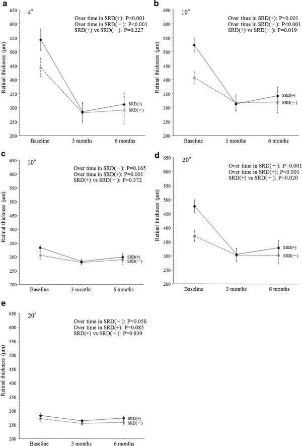
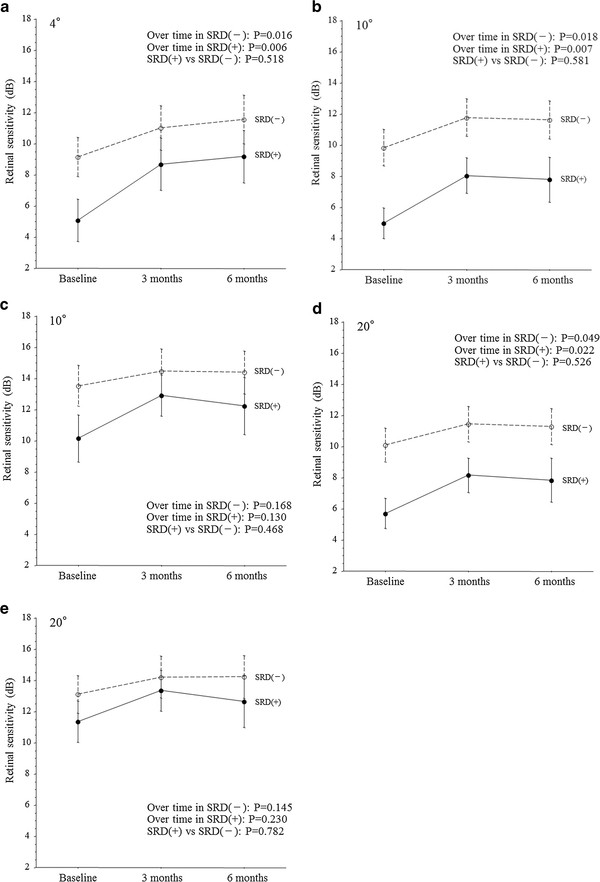
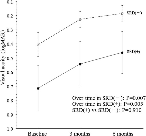
Twenty-one eyes of 21 BRVO patients with macular edema received IVTA. Patients were divided into two groups by optical coherence tomography findings: 11 patients who had cystoid macular edema (CME) with SRD (SRD (+) group) and 10 patients who had CME without SRD (SRD (-) group). Microperimetry was performed with a Micro Perimeter 1 before and at 3 and 6 months after IVTA. Macular thickness was measured by optical coherence tomography. We exchanged the superior and inferior regions to separate the regions into those with and without occlusion. As a result, the superior region was always the occluded region and the inferior region was non-occluded.
In both the SRD (-) group and the SRD (+) group, the mean macular thickness within the central 4° field and the 10° and 20° fields of the occluded region decreased significantly from baseline to 3 and 6 months after IVTA (all P <0.01). Visual acuity also improved significantly in both groups from baseline to 3 and 6 months after IVTA (both P <0.05). In both groups, the mean macular sensitivity (measured with by microperimetry) within the central 4° field and the 10° and 20° fields of the occluded region showed a significant increase from baseline to 3 and 6 months after IVTA (all P <0.05). The trend profiles of macular thickness within the 10° and 20° fields of the occluded region showed significant differences, but there were no significant differences with respect to the trend profiles of visual acuity and macular sensitivity within the central 4° field and the 10° and 20° fields of the occluded region.
These results suggest that IVTA may achieve more marked improvement of macular morphology in BRVO patients with SRD than in those without SRD, while this therapy may have a similar effect on macular function in BRVO patients with or without SRD.
在伴有分支静脉阻塞(BRVO)的黄斑水肿患者中,玻璃体内注射曲安奈德(IVTA)后,视网膜脱离(SRD)对视力、黄斑敏感性和黄斑厚度的影响尚不清楚。
21 例 BRVO 伴黄斑水肿患者的 21 只眼接受 IVTA 治疗。根据光学相干断层扫描(OCT)结果将患者分为两组:11 例合并 SRD 的患者(SRD(+)组)和 10 例无 SRD 的患者(SRD(-)组)。在 IVTA 前、3 个月和 6 个月时使用微视野计进行微视野检查。通过 OCT 测量黄斑厚度。我们交换上下区域,将区域分为闭塞区和非闭塞区。结果,上区域始终是闭塞区,下区域是非闭塞区。
在 SRD(-)组和 SRD(+)组中,闭塞区中央 4°和 10°、20°区域的黄斑平均厚度均从基线至 IVTA 后 3 个月和 6 个月显著降低(均 P<0.01)。两组视力均从基线至 IVTA 后 3 个月和 6 个月显著提高(均 P<0.05)。两组患者的闭塞区中央 4°和 10°、20°区域的黄斑平均敏感度(微视野计测量)均从基线至 IVTA 后 3 个月和 6 个月显著升高(均 P<0.05)。虽然闭塞区 10°和 20°区域的黄斑厚度趋势曲线存在显著差异,但在中央 4°区域和闭塞区 10°和 20°区域的视力和黄斑敏感度的趋势曲线无显著差异。
这些结果表明,IVTA 可能会使伴有 SRD 的 BRVO 患者的黄斑形态改善更为显著,而对伴有或不伴有 SRD 的 BRVO 患者的黄斑功能可能具有相似的作用。