• 文献检索
  • 文档翻译
  • 深度研究
  • 学术资讯
  • Suppr Zotero 插件Zotero 插件
  • 邀请有礼
  • 套餐&价格
  • 历史记录
应用&插件
Suppr Zotero 插件Zotero 插件浏览器插件Mac 客户端Windows 客户端微信小程序
定价
高级版会员购买积分包购买API积分包
服务
文献检索文档翻译深度研究API 文档MCP 服务
关于我们
关于 Suppr公司介绍联系我们用户协议隐私条款
关注我们

Suppr 超能文献

核心技术专利:CN118964589B侵权必究
粤ICP备2023148730 号-1Suppr @ 2026

文献检索

告别复杂PubMed语法,用中文像聊天一样搜索,搜遍4000万医学文献。AI智能推荐,让科研检索更轻松。

立即免费搜索

文件翻译

保留排版,准确专业,支持PDF/Word/PPT等文件格式,支持 12+语言互译。

免费翻译文档

深度研究

AI帮你快速写综述,25分钟生成高质量综述,智能提取关键信息,辅助科研写作。

立即免费体验

一种用于研究动脉粥样硬化斑块纤维帽中胶原蛋白与脂质比例的荧光寿命成像分类方法。

A fluorescence lifetime imaging classification method to investigate the collagen to lipid ratio in fibrous caps of atherosclerotic plaque.

作者信息

Phipps Jennifer E, Sun Yinghua, Fishbein Michael C, Marcu Laura

机构信息

Department of Biomedical Engineering, University of California Davis, Davis, California 95616, USA.

出版信息

Lasers Surg Med. 2012 Sep;44(7):564-71. doi: 10.1002/lsm.22059. Epub 2012 Aug 6.

DOI:10.1002/lsm.22059
PMID:22886522
原文链接:https://pmc.ncbi.nlm.nih.gov/articles/PMC4083183/
Abstract

BACKGROUND AND OBJECTIVE

This study describes a novel fluorescence lifetime imaging (FLIM) classification method to determine the ratio of collagen to lipid content in the fibrous cap of atherosclerotic plaques. Additionally, an analytical process to assess risk of plaque rupture based on this ratio is proposed. Collagen to lipid ratio has been shown to be an important parameter to evaluate structural integrity of the fibrous cap. FLIM and other time-resolved fluorescence techniques have recently been applied to the study of atherosclerosis based on the ability to assess biochemical composition.

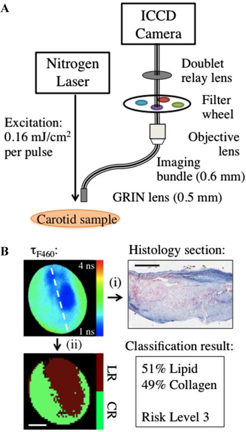
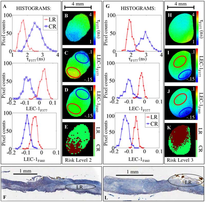
STUDY DESIGN/MATERIALS AND METHODS: Autofluorescence of specimens retrieved during carotid endarterectomy procedures was measured through three optical filters, F377: 377/50 nm, F460: 460/66 nm, and F510: 510/84 nm (center wavelength/bandwidth). A Laguerre deconvolution technique was used for the evaluation of fluorescence decay dynamics. The resulting decay parameters (average fluorescence lifetime and 4 Laguerre coefficients at each of the recorded bandwidths) were used for sample characterization. Linear discriminant analysis (LDA) was used to classify each image into collagen or lipid-rich regions based on these parameters. Ultimately, a risk-level was assigned based on the ratio of collagen to lipid on the surface of the fibrous cap.

RESULTS

FLIM images were acquired in 18 carotid plaque specimens at 43 locations. Classification of collagen and lipid-rich regions within the fibrous cap was performed with sensitivity and specificity of 80% and 82%, respectively.

CONCLUSIONS

Results from this study show that an LDA method of classifying regions of FLIM images of carotid plaque into collagen and lipid-rich regions is capable of being automated and used to rate the risk of plaque rupture based on autofluorescence decay dynamics and without the need for fluorescence intensity or contrast agents.

摘要

背景与目的

本研究描述了一种新型荧光寿命成像(FLIM)分类方法,用于确定动脉粥样硬化斑块纤维帽中胶原蛋白与脂质含量的比例。此外,还提出了一种基于该比例评估斑块破裂风险的分析流程。胶原蛋白与脂质的比例已被证明是评估纤维帽结构完整性的重要参数。基于评估生化成分的能力,FLIM和其他时间分辨荧光技术最近已应用于动脉粥样硬化的研究。

研究设计/材料与方法:通过三个光学滤光片测量颈动脉内膜切除术过程中获取的标本的自发荧光,滤光片分别为F377:377/50nm、F460:460/66nm和F510:510/84nm(中心波长/带宽)。采用拉盖尔反卷积技术评估荧光衰减动力学。所得衰减参数(平均荧光寿命以及每个记录带宽处的4个拉盖尔系数)用于样本表征。基于这些参数,使用线性判别分析(LDA)将每个图像分类为富含胶原蛋白或脂质的区域。最终,根据纤维帽表面胶原蛋白与脂质的比例确定风险等级。

结果

在18个颈动脉斑块标本的43个位置采集了FLIM图像。对纤维帽内富含胶原蛋白和脂质的区域进行分类,灵敏度和特异性分别为80%和82%。

结论

本研究结果表明,一种将颈动脉斑块FLIM图像区域分类为富含胶原蛋白和脂质区域的LDA方法能够实现自动化,并用于基于自发荧光衰减动力学对斑块破裂风险进行评级,且无需荧光强度或造影剂。

https://cdn.ncbi.nlm.nih.gov/pmc/blobs/d5d5/4083183/fd94daa41e1c/nihms-599887-f0002.jpg
https://cdn.ncbi.nlm.nih.gov/pmc/blobs/d5d5/4083183/a94ff92deae3/nihms-599887-f0001.jpg
https://cdn.ncbi.nlm.nih.gov/pmc/blobs/d5d5/4083183/fd94daa41e1c/nihms-599887-f0002.jpg
https://cdn.ncbi.nlm.nih.gov/pmc/blobs/d5d5/4083183/a94ff92deae3/nihms-599887-f0001.jpg
https://cdn.ncbi.nlm.nih.gov/pmc/blobs/d5d5/4083183/fd94daa41e1c/nihms-599887-f0002.jpg

相似文献

1
A fluorescence lifetime imaging classification method to investigate the collagen to lipid ratio in fibrous caps of atherosclerotic plaque.一种用于研究动脉粥样硬化斑块纤维帽中胶原蛋白与脂质比例的荧光寿命成像分类方法。
Lasers Surg Med. 2012 Sep;44(7):564-71. doi: 10.1002/lsm.22059. Epub 2012 Aug 6.
2
Fluorescence lifetime imaging for the characterization of the biochemical composition of atherosclerotic plaques.荧光寿命成像用于动脉粥样硬化斑块生化成分的特征描述。
J Biomed Opt. 2011 Sep;16(9):096018. doi: 10.1117/1.3626865.
3
Biochemical characterization of atherosclerotic plaques by endogenous multispectral fluorescence lifetime imaging microscopy.应用内源性多光谱荧光寿命成像显微镜对动脉粥样硬化斑块进行生化特征分析。
Atherosclerosis. 2012 Feb;220(2):394-401. doi: 10.1016/j.atherosclerosis.2011.10.034. Epub 2011 Nov 15.
4
An Optical Coherence Tomography Assessment of Stent Strut Apposition Based on the Presence of Lipid-Rich Plaque in the Carotid Artery.基于颈动脉富含脂质斑块存在情况的支架支柱贴壁光学相干断层扫描评估
J Endovasc Ther. 2015 Dec;22(6):942-9. doi: 10.1177/1526602815610116. Epub 2015 Oct 13.
5
Label-Free Characterization of Atherosclerotic Plaques Via High-Resolution Multispectral Fluorescence Lifetime Imaging Microscopy.基于高分辨率多光谱荧光寿命成像显微镜的无标记动脉粥样硬化斑块特征分析。
Arterioscler Thromb Vasc Biol. 2023 Jul;43(7):1295-1307. doi: 10.1161/ATVBAHA.123.319339. Epub 2023 May 18.
6
Fluorescence Lifetime Imaging Combined with Conventional Intravascular Ultrasound for Enhanced Assessment of Atherosclerotic Plaques: an Ex Vivo Study in Human Coronary Arteries.荧光寿命成像结合传统血管内超声用于增强动脉粥样硬化斑块评估:人体冠状动脉的体外研究
J Cardiovasc Transl Res. 2015 Jun;8(4):253-63. doi: 10.1007/s12265-015-9627-3. Epub 2015 May 1.
7
Detection of rupture-prone atherosclerotic plaques by time-resolved laser-induced fluorescence spectroscopy.通过时间分辨激光诱导荧光光谱法检测易破裂动脉粥样硬化斑块
Atherosclerosis. 2009 May;204(1):156-64. doi: 10.1016/j.atherosclerosis.2008.08.035. Epub 2008 Sep 6.
8
Characterization of fracture behavior of human atherosclerotic fibrous caps using a miniature single edge notched tensile test.使用微型单边切口拉伸试验对人类动脉粥样硬化纤维帽的断裂行为进行表征。
Acta Biomater. 2016 Oct 1;43:101-111. doi: 10.1016/j.actbio.2016.07.027. Epub 2016 Jul 16.
9
Data Processing Pipeline for Lipid Profiling of Carotid Atherosclerotic Plaque with Mass Spectrometry Imaging.基于质谱成像的颈动脉粥样硬化斑块脂质组学分析的数据处理流程。
J Am Soc Mass Spectrom. 2019 Sep;30(9):1790-1800. doi: 10.1007/s13361-019-02254-y. Epub 2019 Jun 27.
10
Fluorescence lifetime imaging microscopy for the characterization of atherosclerotic plaques.用于表征动脉粥样硬化斑块的荧光寿命成像显微镜技术
Proc SPIE Int Soc Opt Eng. 2009;7161:71612G. doi: 10.1117/12.813087.

引用本文的文献

1
Optical Microscopy and the Extracellular Matrix Structure: A Review.光学显微镜与细胞外基质结构:综述。
Cells. 2021 Jul 12;10(7):1760. doi: 10.3390/cells10071760.
2
Mesoscopic fluorescence lifetime imaging: Fundamental principles, clinical applications and future directions.介观荧光寿命成像:基本原理、临床应用及未来方向。
J Biophotonics. 2021 Jun;14(6):e202000472. doi: 10.1002/jbio.202000472. Epub 2021 Mar 29.
3
Current Advances in the Diagnostic Imaging of Atherosclerosis: Insights into the Pathophysiology of Vulnerable Plaque.

本文引用的文献

1
Multispectral scanning time-resolved fluorescence spectroscopy (TRFS) technique for intravascular diagnosis.用于血管内诊断的多光谱扫描时间分辨荧光光谱(TRFS)技术。
Biomed Opt Express. 2012 Jul 1;3(7):1521-33. doi: 10.1364/BOE.3.001521. Epub 2012 Jun 6.
2
Biochemical characterization of atherosclerotic plaques by endogenous multispectral fluorescence lifetime imaging microscopy.应用内源性多光谱荧光寿命成像显微镜对动脉粥样硬化斑块进行生化特征分析。
Atherosclerosis. 2012 Feb;220(2):394-401. doi: 10.1016/j.atherosclerosis.2011.10.034. Epub 2011 Nov 15.
3
Intra-arterial catheter for simultaneous microstructural and molecular imaging in vivo.
动脉粥样硬化的诊断影像学新进展:易损斑块的病理生理学见解。
Int J Mol Sci. 2020 Apr 23;21(8):2992. doi: 10.3390/ijms21082992.
4
Comprehensive intravascular imaging of atherosclerotic plaque in vivo using optical coherence tomography and fluorescence lifetime imaging.采用光学相干断层扫描和荧光寿命成像技术对活体动脉粥样硬化斑块进行全面的血管内成像。
Sci Rep. 2018 Sep 28;8(1):14561. doi: 10.1038/s41598-018-32951-9.
5
Scanning Acoustic Microscopy and Time-Resolved Fluorescence Spectroscopy for Characterization of Atherosclerotic Plaques.扫描声学显微镜和时间分辨荧光光谱术用于动脉粥样硬化斑块的特征分析。
Sci Rep. 2018 Sep 26;8(1):14378. doi: 10.1038/s41598-018-32788-2.
6
Imaging the Cardiac Extracellular Matrix.心脏细胞外基质的影像学检查
Adv Exp Med Biol. 2018;1098:21-44. doi: 10.1007/978-3-319-97421-7_2.
7
Label-Free Assessment of Collagenase Digestion on Bovine Pericardium Properties by Fluorescence Lifetime Imaging.荧光寿命成像技术对牛心包胶原酶消化特性的无标记评估。
Ann Biomed Eng. 2018 Nov;46(11):1870-1881. doi: 10.1007/s10439-018-2087-6. Epub 2018 Jul 12.
8
Comparing Raman and fluorescence lifetime spectroscopy from human atherosclerotic lesions using a bimodal probe.使用双峰探针比较人动脉粥样硬化病变的拉曼光谱和荧光寿命光谱。
J Biophotonics. 2016 Sep;9(9):958-66. doi: 10.1002/jbio.201500341. Epub 2016 Mar 22.
9
Imaging Fibrosis and Separating Collagens using Second Harmonic Generation and Phasor Approach to Fluorescence Lifetime Imaging.利用二次谐波产生和荧光寿命成像的相量方法对纤维化进行成像并区分胶原蛋白。
Sci Rep. 2015 Aug 21;5:13378. doi: 10.1038/srep13378.
10
Fluorescence Lifetime Imaging Combined with Conventional Intravascular Ultrasound for Enhanced Assessment of Atherosclerotic Plaques: an Ex Vivo Study in Human Coronary Arteries.荧光寿命成像结合传统血管内超声用于增强动脉粥样硬化斑块评估:人体冠状动脉的体外研究
J Cardiovasc Transl Res. 2015 Jun;8(4):253-63. doi: 10.1007/s12265-015-9627-3. Epub 2015 May 1.
体内同时进行微观结构和分子成像的动脉内导管。
Nat Med. 2011 Nov 6;17(12):1680-4. doi: 10.1038/nm.2555.
4
Fluorescence lifetime imaging for the characterization of the biochemical composition of atherosclerotic plaques.荧光寿命成像用于动脉粥样硬化斑块生化成分的特征描述。
J Biomed Opt. 2011 Sep;16(9):096018. doi: 10.1117/1.3626865.
5
A fluorescence lifetime spectroscopy study of matrix metalloproteinases-2 and -9 in human atherosclerotic plaque.基质金属蛋白酶-2 和 -9 在人动脉粥样硬化斑块中的荧光寿命光谱研究。
J Biophotonics. 2011 Sep;4(9):650-8. doi: 10.1002/jbio.201100042. Epub 2011 Jul 19.
6
Imaging the vulnerable plaque.易损斑块的影像学检测
J Am Coll Cardiol. 2011 May 17;57(20):1961-79. doi: 10.1016/j.jacc.2011.02.018.
7
Dynamic tissue analysis using time- and wavelength-resolved fluorescence spectroscopy for atherosclerosis diagnosis.使用时间分辨和波长分辨荧光光谱法进行动态组织分析以诊断动脉粥样硬化
Opt Express. 2011 Feb 28;19(5):3890-901. doi: 10.1364/OE.19.003890.
8
A dual-modality optical coherence tomography and fluorescence lifetime imaging microscopy system for simultaneous morphological and biochemical tissue characterization.一种用于同时进行形态学和生化组织表征的双模态光学相干断层扫描和荧光寿命成像显微镜系统。
Biomed Opt Express. 2010 Jul 16;1(1):186-200. doi: 10.1364/BOE.1.000186.
9
Unreliable assessment of necrotic core by virtual histology intravascular ultrasound in porcine coronary artery disease.猪冠状动脉疾病中虚拟组织学血管内超声对坏死核心评估不可靠。
Circ Cardiovasc Imaging. 2010 Jul;3(4):384-91. doi: 10.1161/CIRCIMAGING.109.919357. Epub 2010 May 11.
10
Intravascular near-infrared fluorescence molecular imaging of atherosclerosis: toward coronary arterial visualization of biologically high-risk plaques.动脉粥样硬化的血管内近红外荧光分子成像:实现对具有高生物学风险斑块的冠状动脉可视化。
J Biomed Opt. 2010 Jan-Feb;15(1):011107. doi: 10.1117/1.3280282.