Bogazici University, Physics Department, Istanbul, 34342, Turkey.
Mehmet Akif Ersoy Thoracic and Cardiovascular Surgery Training Research Hospital, Istanbul, 34303, Turkey.
Sci Rep. 2018 Sep 26;8(1):14378. doi: 10.1038/s41598-018-32788-2.
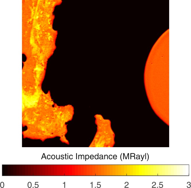
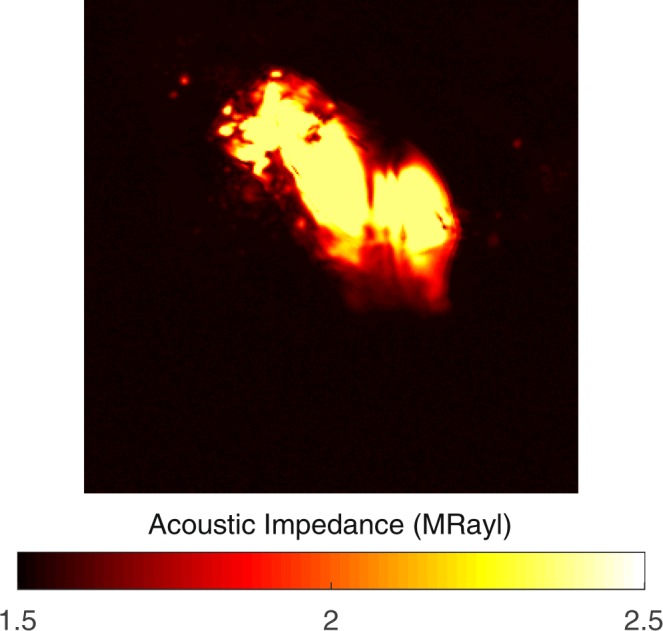
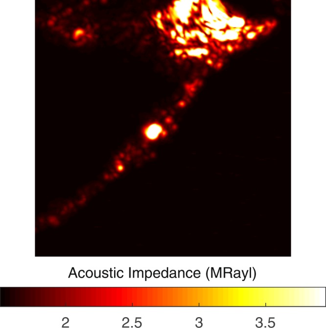
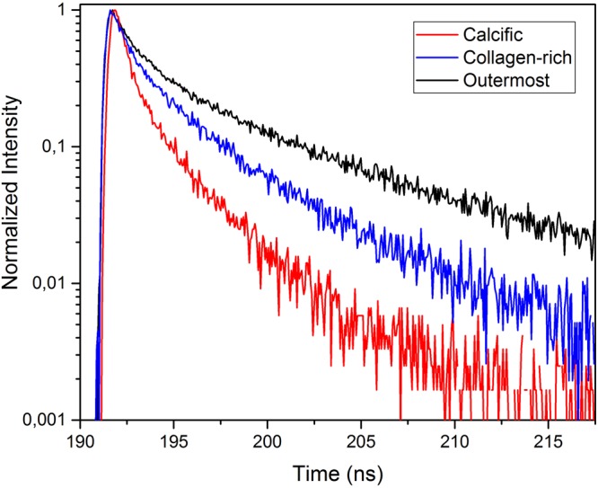
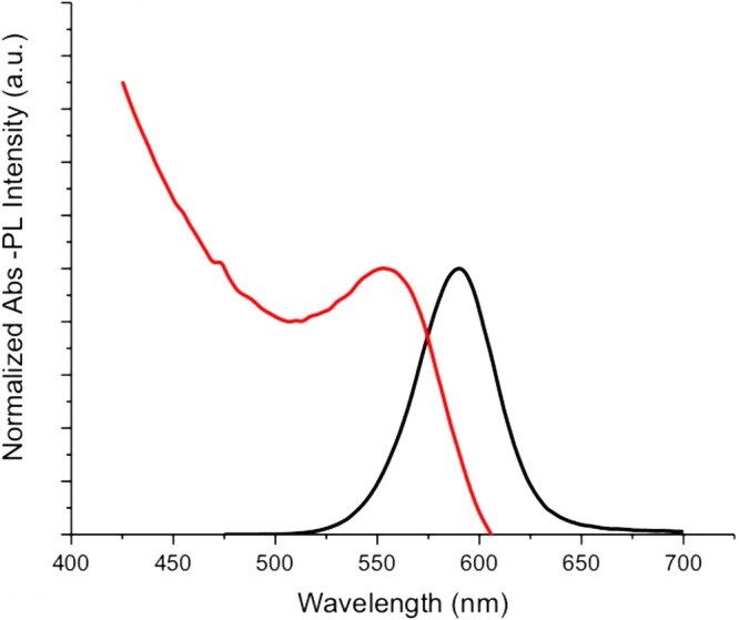
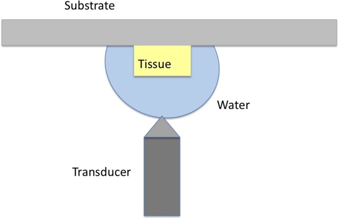
Atherosclerotic plaques constitute the primary cause of heart attack and stroke. However, we still lack a clear identification of the plaques. Here, we evaluate the feasibility of scanning acoustic microscopy (SAM) and time-resolved fluorescence spectroscopy (TRFS) in atherosclerotic plaque characterization. We perform dual-modality microscopic imaging of the human carotid atherosclerotic plaques. We first show that the acoustic impedance values are statistically higher in calcified regions compared with the collagen-rich areas. We then use CdTe/CdS quantum dots for imaging the atherosclerotic plaques by TRFS and show that fluorescence lifetime values of the quantum dots in collagen-rich areas are notably different from the ones in calcified areas. In summary, both modalities are successful in differentiating the calcified regions from the collagen-rich areas within the plaques indicating that these techniques are confirmatory and may be combined to characterize atherosclerotic plaques in the future.
动脉粥样硬化斑块是心脏病发作和中风的主要原因。然而,我们仍然无法明确识别这些斑块。在这里,我们评估扫描声学显微镜(SAM)和时间分辨荧光光谱(TRFS)在动脉粥样硬化斑块特征描述中的可行性。我们对人颈动脉粥样硬化斑块进行双模式微观成像。我们首先表明,与富含胶原蛋白的区域相比,钙化区域的声阻抗值在统计学上更高。然后,我们使用 CdTe/CdS 量子点通过 TRFS 对动脉粥样硬化斑块进行成像,并表明富含胶原蛋白区域的量子点的荧光寿命值与钙化区域的荧光寿命值明显不同。总之,这两种方式都成功地将斑块中的钙化区域与富含胶原蛋白的区域区分开来,表明这些技术具有确认性,将来可能会结合起来对动脉粥样硬化斑块进行特征描述。