NinePoint Medical, One Kendall Square B7501, Cambridge, MA 02139, USA.
Diagn Pathol. 2012 Aug 13;7:98. doi: 10.1186/1746-1596-7-98.
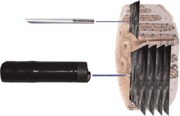
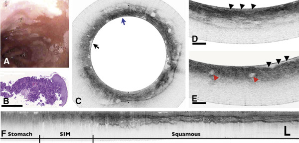
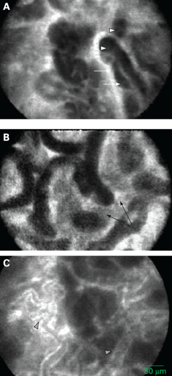
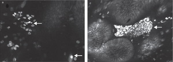
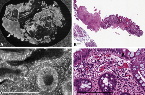
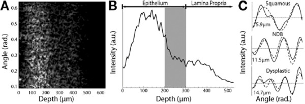
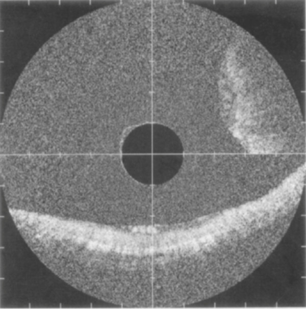
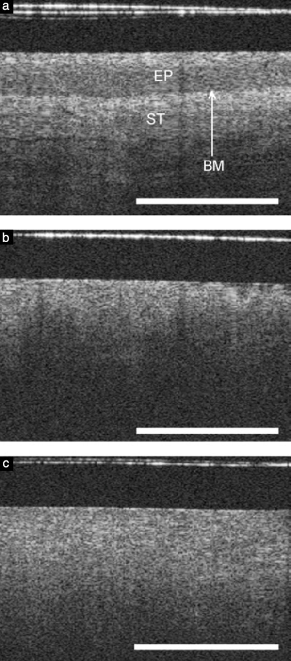
Epithelial cancers account for substantial mortality and are an important public health concern. With the need for earlier detection and treatment of these malignancies, the ability to accurately detect precancerous lesions has an increasingly important role in controlling cancer incidence and mortality. New optical technologies are capable of identifying early pathology in tissues or organs in which cancer is known to develop through stages of dysplasia, including the esophagus, colon, pancreas, liver, bladder, and cervix. These diagnostic imaging advances, together as a field known as optical endomicroscopy, are based on confocal microscopy, spectroscopy-based imaging, and optical coherence tomography (OCT), and function as "optical biopsies," enabling tissue pathology to be imaged in situ and in real time without the need to excise and process specimens as in conventional biopsy and histopathology. Optical biopsy techniques can acquire high-resolution, cross-sectional images of tissue structure on the micron scale through the use of endoscopes, catheters, laparoscopes, and needles. Since the inception of these technologies, dramatic technological advances in accuracy, speed, and functionality have been realized. The current paradigm of optical biopsy, or single-area, point-based images, is slowly shifting to more comprehensive microscopy of larger tracts of mucosa. With the development of Fourier-domain OCT, also known as optical frequency domain imaging or, more recently, volumetric laser endomicroscopy, comprehensive surveillance of the entire distal esophagus is now achievable at speeds that were not possible with conventional OCT technologies. Optical diagnostic technologies are emerging as clinically useful tools with the potential to set a new standard for real-time diagnosis. New imaging techniques enable visualization of high-resolution, cross-sectional images and offer the opportunity to guide biopsy, allowing maximal diagnostic yields and appropriate staging without the limitations and risks inherent with current random biopsy protocols. However, the ability of these techniques to achieve widespread adoption in clinical practice depends on future research designed to improve accuracy and allow real-time data transmission and storage, thereby linking pathology to the treating physician. These imaging advances are expected to eventually offer a see-and-treat paradigm, leading to improved patient care and potential cost reduction.
The virtual slide(s) for this article can be found here: http://www.diagnosticpathology.diagnomx.eu/vs/5372548637202968.
上皮性癌导致大量死亡,是一个重要的公共卫生关注点。随着对这些恶性肿瘤进行更早检测和治疗的需求,准确检测癌前病变在控制癌症发病率和死亡率方面的作用日益重要。新的光学技术能够识别已知在组织或器官中发展的癌症的早期病理学,这些组织或器官经历了包括食管、结肠、胰腺、肝脏、膀胱和子宫颈等部位的异型增生阶段。这些诊断成像方面的进步,统称为光学内窥技术,基于共焦显微镜、基于光谱的成像和光相干断层扫描(OCT),作为“光学活检”,能够在无需切除和处理标本的情况下原位实时成像组织病理学,就像传统活检和组织病理学那样。光学活检技术可以通过使用内窥镜、导管、腹腔镜和针,获取组织结构的高分辨率、横截面微米级图像。自从这些技术问世以来,在准确性、速度和功能方面取得了巨大的技术进步。目前的光学活检范例,或者说是单点、基于区域的图像,正在逐渐转向更大面积的黏膜综合显微镜。随着傅里叶域 OCT(也称为光频域成像,或最近的容积激光内窥技术)的发展,整个远端食管的全面监测现在可以以传统 OCT 技术无法实现的速度进行。光学诊断技术正在成为具有临床实用价值的工具,有可能为实时诊断设定新标准。新的成像技术能够可视化高分辨率、横截面图像,并提供引导活检的机会,在不局限于当前随机活检方案的固有局限性和风险的情况下,实现最大的诊断效果和适当的分期。然而,这些技术在临床实践中广泛采用的能力取决于旨在提高准确性并允许实时数据传输和存储的未来研究,从而将病理学与治疗医生联系起来。这些成像方面的进步有望最终提供一种“观察和治疗”的范例,从而改善患者护理并降低潜在成本。
本文的虚拟幻灯片可在此处找到:http://www.diagnosticpathology.diagnomx.eu/vs/5372548637202968。