Department of Radiology, University of Washington, Seattle, WA, USA.
JACC Cardiovasc Imaging. 2012 Aug;5(8):798-804. doi: 10.1016/j.jcmg.2012.03.014.
This study sought to determine the immediate and long-term effects of intraplaque hemorrhage (IPH) on plaque progression in the carotid artery.
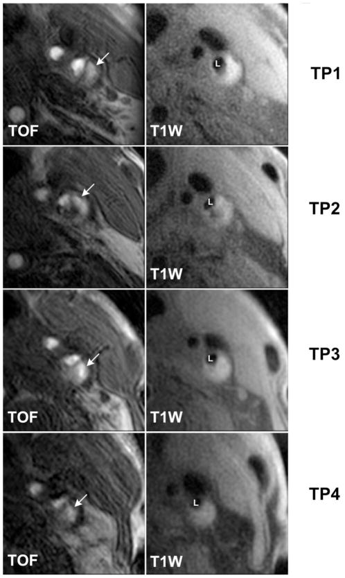
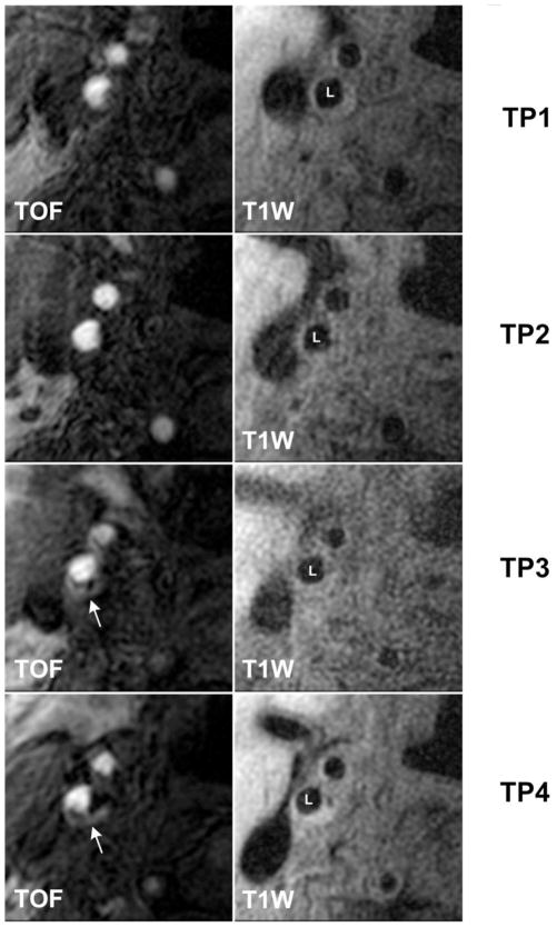
Previous studies have associated IPH in the carotid artery with more rapid plaque progression. However, the time course and long-term effect remain unknown. Carotid magnetic resonance imaging is a noninvasive imaging technique that has been validated with histology for the accurate in vivo detection of IPH and measurement of plaque burden.
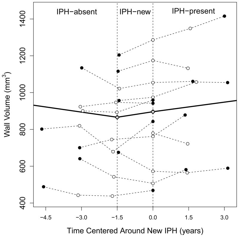
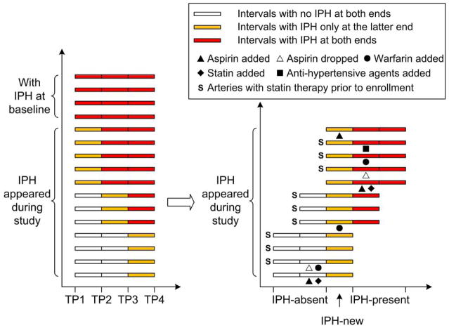
Asymptomatic subjects with 50% to 79% carotid stenosis underwent carotid magnetic resonance imaging at baseline and then serially every 18 months for a total of 54 months. Subjects with IPH present in at least 1 carotid artery at 54 months were selected. Subsequently, presence/absence of IPH and wall volume were determined independently in all time points for both sides. A piece-wise progression curve was fit by using a linear mixed model to compare progression rates described as annualized changes in wall volume between periods defined by their relationship to IPH development.
From 14 subjects who exhibited IPH at 54 months, 12 arteries were found to have developed IPH during the study period. The progression rates were -20.5 ± 13.1, 20.5 ± 13.6, and 16.5 ± 10.8 mm(3)/year before, during, and after IPH development, respectively. The progression rate during IPH development tended to be higher than the period before (p = 0.080) but comparable to the period after (p = 0.845). The progression rate in the combined period during/after IPH development was 18.3 ± 6.5 mm(3)/year, which indicated significant progression (p = 0.008 compared with a slope of 0) and was higher than the period before IPH development (p = 0.018). No coincident ischemic events were noted for new IPH.
The development of IPH posed an immediate and long-term promoting effect on plaque progression. IPH seems to alter the biology and natural history of carotid atherosclerosis. Early identification of patients with IPH may prove invaluable in optimizing management to minimize future sequelae.
本研究旨在确定斑块内出血(IPH)对颈动脉斑块进展的即刻和长期影响。
先前的研究表明,颈动脉中的 IPH 与斑块进展更快有关。然而,其时间过程和长期影响尚不清楚。颈动脉磁共振成像(MRI)是一种非侵入性成像技术,已通过组织学验证,可准确地在体内检测 IPH 并测量斑块负担。
无症状的颈动脉狭窄程度为 50%至 79%的患者在基线时进行颈动脉 MRI 检查,然后每 18 个月进行一次连续检查,共 54 个月。选择在 54 个月时至少有 1 条颈动脉存在 IPH 的患者。随后,在所有时间点独立确定双侧 IPH 的存在/不存在和壁体积。使用线性混合模型拟合分段进展曲线,以比较根据与 IPH 发展关系定义的时间段描述的壁体积年变化率,从而比较进展率。
在 54 个月时表现出 IPH 的 14 名患者中,有 12 条动脉在研究期间发展出了 IPH。进展率分别为 -20.5±13.1、20.5±13.6 和 16.5±10.8mm³/年,在 IPH 发展之前、期间和之后。IPH 发展期间的进展率倾向于高于之前的时期(p=0.080),但与之后的时期相当(p=0.845)。IPH 发展期间/之后的联合时期的进展率为 18.3±6.5mm³/年,表明明显进展(与斜率为 0 相比,p=0.008),高于 IPH 发展之前的时期(p=0.018)。新的 IPH 没有并发缺血性事件。
IPH 的发展对斑块进展有即刻和长期的促进作用。IPH 似乎改变了颈动脉粥样硬化的生物学和自然史。早期识别存在 IPH 的患者可能对优化管理以尽量减少未来的后遗症具有重要意义。