• 文献检索
  • 文档翻译
  • 深度研究
  • 学术资讯
  • Suppr Zotero 插件Zotero 插件
  • 邀请有礼
  • 套餐&价格
  • 历史记录
应用&插件
Suppr Zotero 插件Zotero 插件浏览器插件Mac 客户端Windows 客户端微信小程序
定价
高级版会员购买积分包购买API积分包
服务
文献检索文档翻译深度研究API 文档MCP 服务
关于我们
关于 Suppr公司介绍联系我们用户协议隐私条款
关注我们

Suppr 超能文献

核心技术专利:CN118964589B侵权必究
粤ICP备2023148730 号-1Suppr @ 2026

文献检索

告别复杂PubMed语法,用中文像聊天一样搜索,搜遍4000万医学文献。AI智能推荐,让科研检索更轻松。

立即免费搜索

文件翻译

保留排版,准确专业,支持PDF/Word/PPT等文件格式,支持 12+语言互译。

免费翻译文档

深度研究

AI帮你快速写综述,25分钟生成高质量综述,智能提取关键信息,辅助科研写作。

立即免费体验

肌动蛋白封端蛋白通过拮抗 mDia1 的肌动蛋白活性促进微管稳定性。

Actin-capping protein promotes microtubule stability by antagonizing the actin activity of mDia1.

机构信息

Department of Pathology and Cell Biology, Columbia University, New York, NY 10032, USA.

出版信息

Mol Biol Cell. 2012 Oct;23(20):4032-40. doi: 10.1091/mbc.E12-05-0338. Epub 2012 Aug 23.

DOI:10.1091/mbc.E12-05-0338
PMID:22918941
原文链接:https://pmc.ncbi.nlm.nih.gov/articles/PMC3469518/
Abstract

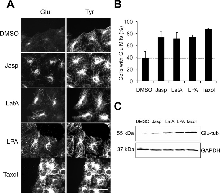
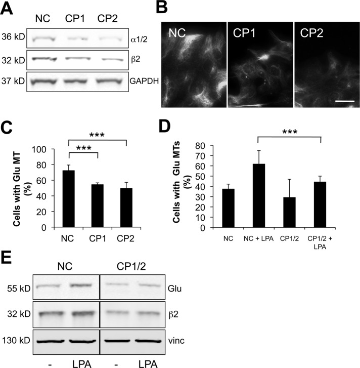
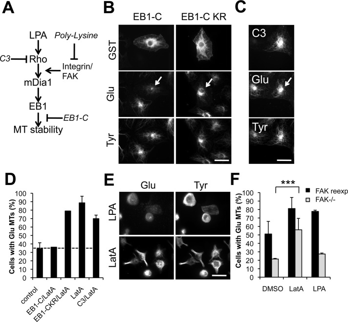
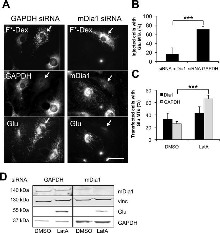
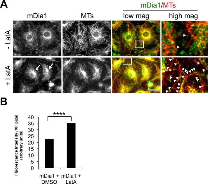
In migrating fibroblasts, RhoA and its effector mDia1 regulate the selective stabilization of microtubules (MTs) polarized in the direction of migration. The conserved formin homology 2 domain of mDia1 is involved both in actin polymerization and MT stabilization, and the relationship between these two activities is unknown. We found that latrunculin A (LatA) and jasplakinolide, actin drugs that release mDia1 from actin filament barbed ends, stimulated stable MT formation in serum-starved fibroblasts and caused a redistribution of mDia1 onto MTs. Knockdown of mDia1 by small interfering RNA (siRNA) prevented stable MT induction by LatA, whereas blocking upstream Rho or integrin signaling had no effect. In search of physiological regulators of mDia1, we found that actin-capping protein induced stable MTs in an mDia1-dependent manner and inhibited the translocation of mDia on the ends of growing actin filaments. Knockdown of capping protein by siRNA reduced stable MT levels in proliferating cells and in starved cells stimulated with lysophosphatidic acid. These results show that actin-capping protein is a novel regulator of MT stability that functions by antagonizing mDia1 activity toward actin filaments and suggest a novel form of actin-MT cross-talk in which a single factor acts sequentially on actin and MTs.

摘要

在迁移的成纤维细胞中,RhoA 和其效应因子 mDia1 调节向迁移方向极化的微管 (MTs) 的选择性稳定。mDia1 的保守的formin 同源 2 结构域既参与肌动蛋白聚合又参与 MT 稳定,而这两种活性之间的关系尚不清楚。我们发现,Latrunculin A (LatA) 和 Jasplakinolide,肌动蛋白药物,可将 mDia1 从肌动蛋白丝的帽状末端释放,刺激血清饥饿的成纤维细胞中稳定的 MT 形成,并导致 mDia1 在 MT 上重新分布。小干扰 RNA (siRNA) 敲低 mDia1 可阻止 LatA 诱导的稳定 MT 诱导,而阻断上游 Rho 或整合素信号无影响。在寻找 mDia1 的生理调节剂时,我们发现肌动蛋白加帽蛋白以 mDia1 依赖的方式诱导稳定的 MT,并抑制 mDia 在生长肌动蛋白纤维末端的易位。siRNA 敲低加帽蛋白可降低增殖细胞和用溶血磷脂酸刺激的饥饿细胞中的稳定 MT 水平。这些结果表明,肌动蛋白加帽蛋白是 MT 稳定性的一种新型调节剂,通过拮抗 mDia1 对肌动蛋白纤维的活性起作用,并提示一种新的肌动蛋白-MT 串扰形式,其中单个因子依次作用于肌动蛋白和 MT。

https://cdn.ncbi.nlm.nih.gov/pmc/blobs/d865/3469518/50ff26fbad97/4032fig6.jpg
https://cdn.ncbi.nlm.nih.gov/pmc/blobs/d865/3469518/8bbcb6ffc000/4032fig1.jpg
https://cdn.ncbi.nlm.nih.gov/pmc/blobs/d865/3469518/15ed4f179d52/4032fig2.jpg
https://cdn.ncbi.nlm.nih.gov/pmc/blobs/d865/3469518/965f982e9e8b/4032fig3.jpg
https://cdn.ncbi.nlm.nih.gov/pmc/blobs/d865/3469518/4fc396ba6e97/4032fig4.jpg
https://cdn.ncbi.nlm.nih.gov/pmc/blobs/d865/3469518/e926a7cd6c36/4032fig5.jpg
https://cdn.ncbi.nlm.nih.gov/pmc/blobs/d865/3469518/50ff26fbad97/4032fig6.jpg

相似文献

1
Actin-capping protein promotes microtubule stability by antagonizing the actin activity of mDia1.肌动蛋白封端蛋白通过拮抗 mDia1 的肌动蛋白活性促进微管稳定性。
Mol Biol Cell. 2012 Oct;23(20):4032-40. doi: 10.1091/mbc.E12-05-0338. Epub 2012 Aug 23.
2
Actin polymerization-driven molecular movement of mDia1 in living cells.活细胞中肌动蛋白聚合驱动的mDia1分子运动。
Science. 2004 Mar 26;303(5666):2007-10. doi: 10.1126/science.1093923.
3
An mDia1-INF2 formin activation cascade facilitated by IQGAP1 regulates stable microtubules in migrating cells.由IQGAP1促进的mDia1-INF2formin激活级联反应调节迁移细胞中的稳定微管。
Mol Biol Cell. 2016 Jun 1;27(11):1797-808. doi: 10.1091/mbc.E15-07-0489. Epub 2016 Mar 30.
4
The formin mDia regulates GSK3beta through novel PKCs to promote microtubule stabilization but not MTOC reorientation in migrating fibroblasts.formin蛋白mDia通过新型蛋白激酶C调节糖原合成酶激酶3β,以促进迁移成纤维细胞中的微管稳定,但不促进微管组织中心重新定向。
Mol Biol Cell. 2006 Dec;17(12):5004-16. doi: 10.1091/mbc.e05-10-0914. Epub 2006 Sep 20.
5
Differential interactions of the formins INF2, mDia1, and mDia2 with microtubules.formin INF2、mDia1 和 mDia2 与微管的差异化相互作用。
Mol Biol Cell. 2011 Dec;22(23):4575-87. doi: 10.1091/mbc.E11-07-0616. Epub 2011 Oct 12.
6
The formin mDia2 stabilizes microtubules independently of its actin nucleation activity.formin蛋白mDia2可独立于其肌动蛋白成核活性来稳定微管。
J Cell Biol. 2008 May 5;181(3):523-36. doi: 10.1083/jcb.200709029.
7
Specificity of interactions between mDia isoforms and Rho proteins.mDia 亚型与 Rho 蛋白之间相互作用的特异性。
J Biol Chem. 2008 Dec 12;283(50):35236-46. doi: 10.1074/jbc.M805634200. Epub 2008 Sep 30.
8
Kif4 interacts with EB1 and stabilizes microtubules downstream of Rho-mDia in migrating fibroblasts.Kif4 与 EB1 相互作用,并稳定 Rho-mDia 下游迁移成纤维细胞中的微管。
PLoS One. 2014 Mar 21;9(3):e91568. doi: 10.1371/journal.pone.0091568. eCollection 2014.
9
Essential and nonredundant roles for Diaphanous formins in cortical microtubule capture and directed cell migration.Diaphanous成膜体在皮质微管捕获和定向细胞迁移中起重要且非冗余的作用。
Mol Biol Cell. 2014 Mar;25(5):658-68. doi: 10.1091/mbc.E13-08-0482. Epub 2014 Jan 8.
10
G-actin regulates rapid induction of actin nucleation by mDia1 to restore cellular actin polymers.球形肌动蛋白(G-肌动蛋白)通过mDia1调节肌动蛋白成核的快速诱导,以恢复细胞中的肌动蛋白聚合物。
J Cell Sci. 2008 Oct 15;121(Pt 20):3403-12. doi: 10.1242/jcs.030940. Epub 2008 Sep 30.

引用本文的文献

1
Tau-Driven Coordination of Microtubule-Actin Crosstalk in Cell-Sized Vesicles.细胞大小囊泡中tau驱动的微管-肌动蛋白串扰协调
bioRxiv. 2025 Aug 13:2025.08.12.669979. doi: 10.1101/2025.08.12.669979.
2
Protrusion-Derived Extracellular Vesicles (PD-EVs) and Their Diverse Origins: Key Players in Cellular Communication, Cancer Progression, and T Cell Modulation.突起衍生的细胞外囊泡(PD-EVs)及其多样起源:细胞通讯、癌症进展和T细胞调节中的关键参与者
Biol Cell. 2025 Jun;117(6):e70018. doi: 10.1111/boc.70018.
3
Mechanical confinement prevents ectopic platelet release.

本文引用的文献

1
Differential interactions of the formins INF2, mDia1, and mDia2 with microtubules.formin INF2、mDia1 和 mDia2 与微管的差异化相互作用。
Mol Biol Cell. 2011 Dec;22(23):4575-87. doi: 10.1091/mbc.E11-07-0616. Epub 2011 Oct 12.
2
The myosin passenger protein Smy1 controls actin cable structure and dynamics by acting as a formin damper.肌球蛋白乘客蛋白 Smy1 通过充当formin 阻尼器来控制肌动蛋白电缆结构和动力学。
Dev Cell. 2011 Aug 16;21(2):217-30. doi: 10.1016/j.devcel.2011.07.004.
3
Aurora B regulates formin mDia3 in achieving metaphase chromosome alignment.
机械束缚可防止血小板异位释放。
Proc Natl Acad Sci U S A. 2024 Sep 17;121(38):e2407829121. doi: 10.1073/pnas.2407829121. Epub 2024 Sep 5.
4
Collective dynamics of actin and microtubule and its crosstalk mediated by FHDC1.肌动蛋白和微管的集体动力学及其由FHDC1介导的串扰。
Front Cell Dev Biol. 2024 Mar 19;11:1261117. doi: 10.3389/fcell.2023.1261117. eCollection 2023.
5
Multicomponent regulation of actin barbed end assembly by twinfilin, formin and capping protein.双胎蛋白、形成蛋白和加帽蛋白对肌动蛋白突状末端组装的多组分调节。
Nat Commun. 2023 Jul 6;14(1):3981. doi: 10.1038/s41467-023-39655-3.
6
Investigating Mammalian Formins with SMIFH2 Fifteen Years in: Novel Targets and Unexpected Biology.用 SMIFH2 研究哺乳动物形成蛋白十五年:新靶点和意外的生物学。
Int J Mol Sci. 2023 May 21;24(10):9058. doi: 10.3390/ijms24109058.
7
Multicomponent regulation of actin barbed end assembly by twinfilin, formin and capping protein.肌动蛋白丝正极组装的多组分调控:双肌动蛋白、formin和封端蛋白的作用
bioRxiv. 2023 Apr 26:2023.04.24.538010. doi: 10.1101/2023.04.24.538010.
8
Dia- and Rok-dependent enrichment of capping proteins in a cortical region.在皮质区域中 Dia 和 Rok 依赖性的盖帽蛋白富集。
J Cell Sci. 2021 Nov 1;134(21). doi: 10.1242/jcs.258973. Epub 2021 Nov 5.
9
Formin Activity and mDia1 Contribute to Maintain Axon Initial Segment Composition and Structure.formin 活性和 mDia1 有助于维持轴突起始段的组成和结构。
Mol Neurobiol. 2021 Dec;58(12):6153-6169. doi: 10.1007/s12035-021-02531-6. Epub 2021 Aug 30.
10
Measuring expression heterogeneity of single-cell cytoskeletal protein complexes.测量单细胞细胞骨架蛋白复合物的表达异质性。
Nat Commun. 2021 Aug 17;12(1):4969. doi: 10.1038/s41467-021-25212-3.
极光 B 调节formin mDia3 以实现中期染色体的排列。
Dev Cell. 2011 Mar 15;20(3):342-52. doi: 10.1016/j.devcel.2011.01.008.
4
Adenomatous polyposis coli protein nucleates actin assembly and synergizes with the formin mDia1.腺瘤性结肠息肉病蛋白促使肌动蛋白组装成核,并与formin蛋白mDia1协同作用。
J Cell Biol. 2010 Jun 28;189(7):1087-96. doi: 10.1083/jcb.201001016. Epub 2010 Jun 21.
5
Formins and microtubules.formin蛋白与微管
Biochim Biophys Acta. 2010 Feb;1803(2):164-73. doi: 10.1016/j.bbamcr.2009.07.006. Epub 2009 Jul 23.
6
Antimitotic chemotherapeutics promote adhesive responses in detached and circulating tumor cells.抗有丝分裂化疗药物促进游离和循环肿瘤细胞的黏附反应。
Breast Cancer Res Treat. 2010 May;121(1):65-78. doi: 10.1007/s10549-009-0457-3. Epub 2009 Jul 11.
7
Displacement of formins from growing barbed ends by bud14 is critical for actin cable architecture and function.Bud14将formin从生长的肌动蛋白丝末端置换出来对肌动蛋白电缆的结构和功能至关重要。
Dev Cell. 2009 Feb;16(2):292-302. doi: 10.1016/j.devcel.2008.12.001.
8
Beyond polymer polarity: how the cytoskeleton builds a polarized cell.超越聚合物极性:细胞骨架如何构建极化细胞。
Nat Rev Mol Cell Biol. 2008 Nov;9(11):860-73. doi: 10.1038/nrm2522.
9
G-actin regulates rapid induction of actin nucleation by mDia1 to restore cellular actin polymers.球形肌动蛋白(G-肌动蛋白)通过mDia1调节肌动蛋白成核的快速诱导,以恢复细胞中的肌动蛋白聚合物。
J Cell Sci. 2008 Oct 15;121(Pt 20):3403-12. doi: 10.1242/jcs.030940. Epub 2008 Sep 30.
10
New insights into mechanism and regulation of actin capping protein.肌动蛋白封端蛋白的机制与调控新见解
Int Rev Cell Mol Biol. 2008;267:183-206. doi: 10.1016/S1937-6448(08)00604-7.