Center for Imaging of Neurodegenerative Diseases, Veterans Affairs Medical Center, San Francisco, CA 94121, USA.
BMC Neurol. 2012 Aug 25;12:83. doi: 10.1186/1471-2377-12-83.
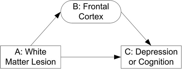
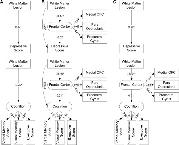
To test the hypothesis that white matter lesions (WML) are primarily associated with regional frontal cortical volumes, and to determine the mediating effects of these regional frontal cortices on the associations of WML with depressive symptoms and cognitive dysfunction.
Structural brains MRIs were performed on 161 participants: cognitively normal, cognitive impaired but not demented, and demented participants. Lobar WML volumes, regional frontal cortical volumes, depressive symptom severity, and cognitive abilities were measured. Multiple linear regression analyses were used to identify WML volume effects on frontal cortical volume. Structural equation modeling was used to determine the MRI-depression and the MRI-cognition path relationships.
WML predicted frontal cortical volume, particularly in medial orbirtofrontal cortex, irrespective of age, gender, education, and group status. WML directly predicted depressive score, and this relationship was not mediated by regional frontal cortices. In contrast, the association between WML and cognitive function was indirect and mediated by regional frontal cortices.
These findings suggest that the neurobiological mechanisms underpinning depressive symptoms and cognitive dysfunction in older adults may differ.
为了检验这样一个假设,即脑白质病变(WML)主要与额皮质区域体积相关,并确定这些额皮质区域在 WML 与抑郁症状和认知功能障碍之间的关联中的中介作用。
对 161 名参与者进行了结构脑 MRI 检查:认知正常、认知障碍但未痴呆和痴呆参与者。测量了脑叶 WML 体积、额皮质区域体积、抑郁症状严重程度和认知能力。采用多元线性回归分析确定 WML 体积对额皮质体积的影响。采用结构方程模型确定 MRI-抑郁和 MRI-认知路径关系。
WML 预测了额皮质体积,尤其是在中线眶额皮质,与年龄、性别、教育程度和组状态无关。WML 直接预测抑郁评分,且该关系不受额皮质区域的影响。相比之下,WML 与认知功能之间的关联是间接的,并受额皮质区域的影响。
这些发现表明,老年人抑郁症状和认知功能障碍的神经生物学机制可能不同。