Lee Christopher Sd, Burnsed Olivia A, Raghuram Vineeth, Kalisvaart Jonathan, Boyan Barbara D, Schwartz Zvi
Stem Cell Res Ther. 2012 Aug 24;3(4):35. doi: 10.1186/scrt126.
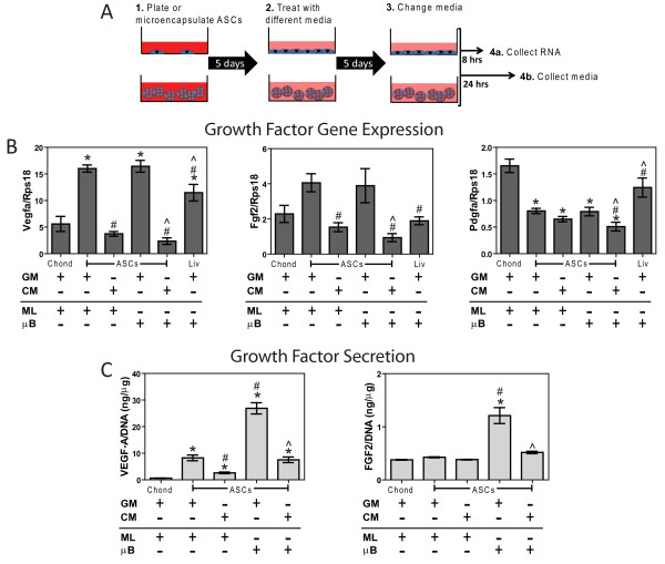
Adipose stem cells (ASCs) secrete many trophic factors that can stimulate tissue repair, including angiogenic factors, but little is known about how ASCs and their secreted factors influence cartilage regeneration. Therefore, the aim of this study was to determine the effects ASC-secreted factors have in repairing chondral defects.
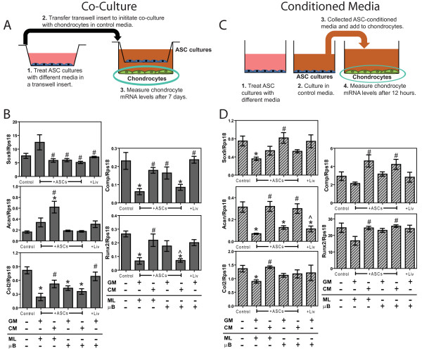
ASCs isolated from male Sprague Dawley rats were cultured in monolayer or alginate microbeads supplemented with growth (GM) or chondrogenic medium (CM). Subsequent co-culture, conditioned media, and in vivo cartilage defect studies were performed.
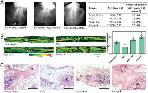
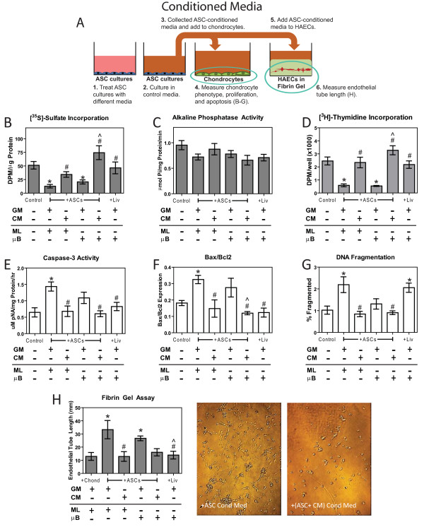
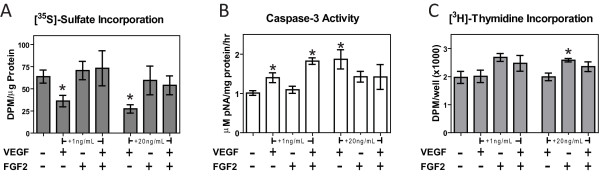
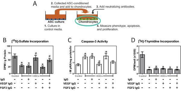
ASC monolayers and microbeads cultured in CM had decreased FGF-2 gene expression and VEGF-A secretion compared to ASCs cultured in GM. Chondrocytes co-cultured with GM-cultured ASCs for 7 days had decreased mRNAs for col2, comp, and runx2. Chondrocytes treated for 12 or 24 hours with conditioned medium from GM-cultured ASCs had reduced sox9, acan, and col2 mRNAs; reduced proliferation and proteoglycan synthesis; and increased apoptosis. ASC-conditioned medium also increased endothelial cell tube lengthening whereas conditioned medium from CM-cultured ASCs had no effect. Treating ASCs with CM reduced or abolished these deleterious effects while adding a neutralizing antibody for VEGF-A eliminated ASC-conditioned medium induced chondrocyte apoptosis and restored proteoglycan synthesis. FGF-2 also mitigated the deleterious effects VEGF-A had on chondrocyte apoptosis and phenotype. When GM-grown ASC pellets were implanted in 1 mm non-critical hyaline cartilage defects in vivo, cartilage regeneration was inhibited as evaluated by radiographic and equilibrium partitioning of an ionic contrast agent via microCT imaging. Histology revealed that defects with GM-cultured ASCs had no tissue ingrowth from the edges of the defect whereas empty defects and defects with CM-grown ASCs had similar amounts of neocartilage formation.
ASCs must be treated to reduce the secretion of VEGF-A and other factors that inhibit cartilage regeneration, which can significantly influence how ASCs are used for repairing hyaline cartilage.
脂肪干细胞(ASC)分泌多种可刺激组织修复的营养因子,包括血管生成因子,但关于ASC及其分泌因子如何影响软骨再生的了解甚少。因此,本研究的目的是确定ASC分泌因子在修复软骨缺损中的作用。
从雄性Sprague Dawley大鼠分离的ASC在补充有生长培养基(GM)或软骨形成培养基(CM)的单层或藻酸盐微珠中培养。随后进行共培养、条件培养基和体内软骨缺损研究。
与在GM中培养的ASC相比,在CM中培养的ASC单层和微珠的FGF-2基因表达和VEGF-A分泌减少。与GM培养的ASC共培养7天的软骨细胞,其col2、comp和runx2的mRNA减少。用GM培养的ASC的条件培养基处理12或24小时的软骨细胞,sox9、acan和col2的mRNA减少;增殖和蛋白聚糖合成减少;凋亡增加。ASC条件培养基还增加了内皮细胞管的延长,而CM培养的ASC的条件培养基则没有作用。用CM处理ASC可减少或消除这些有害影响,而添加VEGF-A中和抗体可消除ASC条件培养基诱导的软骨细胞凋亡并恢复蛋白聚糖合成。FGF-2也减轻了VEGF-A对软骨细胞凋亡和表型的有害影响。当将GM培养的ASC微球植入体内1mm的非关键性透明软骨缺损中时,通过X线摄影和离子造影剂经微CT成像的平衡分配评估,软骨再生受到抑制。组织学显示,GM培养的ASC的缺损处没有从缺损边缘向内生长的组织,而空白缺损和CM培养的ASC的缺损处有相似数量的新软骨形成。
必须对ASC进行处理以减少VEGF-A和其他抑制软骨再生的因子的分泌,这会显著影响ASC用于修复透明软骨的方式。