Radiology Department, Izmir Education and Research Hospital, Izmir, Turkey.
Radiol Oncol. 2010 Jun;44(2):97-102. doi: 10.2478/v10019-010-0006-z. Epub 2010 May 24.
The aim of the study was to investigate the value of diffusion weighted MR imaging in the diagnosis of Modic type 1 change, which may be confused with the acute infectious spondylodiscitis on conventional MR imaging.
Twenty-seven patients with erosive intervertebral osteochondrosis, Modic type 1 and 18 patients with spondylodiscitis were included in this retrospective study. All images were acquired using on 1.5 Tesla MR units. Lumbar spinal MR imaging of 45 patients were retrieved from a digital database of a radiology record system and evaluated by one experienced radiologist. Patients with Modic type 1 change had CT slices obtained from the diseased disc space and the affected vertebrae.
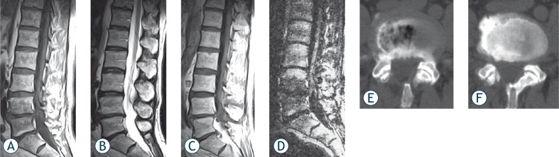
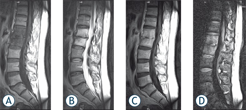
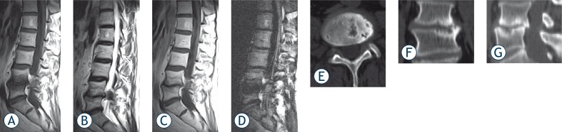
Bone marrow adjacent to the vertebral end plate in both Modic type 1 change and acute spondylodiscitis were hypointense on T1-weighted images. On T2-weighted images corresponding levels of vertebral end-plates showed hyperintense signal intensity in both group. All the patients with spondylodiscitis and Modic type 1 change were hyperintense and hypointense on diffusion-weighted MR images, respectively.
Our findings suggest that diffusion weighted MR imaging is an useful method in differentiating Modic type 1 changes from acute spondylodiscitis, both of which may mimic each other, either on clinical or conventional MRI findings.
本研究旨在探讨弥散加权磁共振成像(DWI)在诊断 Modic 型 1 改变中的价值,因为这种改变在常规磁共振成像(MRI)上可能与急性感染性脊椎炎相混淆。
本回顾性研究纳入了 27 例侵蚀性椎间盘骨软骨炎、Modic 型 1 改变患者和 18 例脊椎炎患者。所有图像均在 1.5T 磁共振仪上采集。从放射学记录系统的数字数据库中检索了 45 例患者的腰椎 MRI,并由一位经验丰富的放射科医生进行评估。Modic 型 1 改变患者在患病椎间盘间隙和受累椎体上均获得 CT 切片。
T1 加权图像上,椎间盘终板旁骨髓在 Modic 型 1 改变和急性脊椎炎中均呈低信号。在 T2 加权图像上,两组相应的椎体终板均呈高信号强度。所有脊椎炎和 Modic 型 1 改变患者的弥散加权磁共振成像(DWI)均呈高信号和低信号。
我们的研究结果表明,弥散加权磁共振成像(DWI)是一种有用的方法,可用于区分 Modic 型 1 改变和急性脊椎炎,因为这两种疾病在临床或常规 MRI 上均可能相互模仿。