• 文献检索
  • 文档翻译
  • 深度研究
  • 学术资讯
  • Suppr Zotero 插件Zotero 插件
  • 邀请有礼
  • 套餐&价格
  • 历史记录
应用&插件
Suppr Zotero 插件Zotero 插件浏览器插件Mac 客户端Windows 客户端微信小程序
定价
高级版会员购买积分包购买API积分包
服务
文献检索文档翻译深度研究API 文档MCP 服务
关于我们
关于 Suppr公司介绍联系我们用户协议隐私条款
关注我们

Suppr 超能文献

核心技术专利:CN118964589B侵权必究
粤ICP备2023148730 号-1Suppr @ 2026

文献检索

告别复杂PubMed语法,用中文像聊天一样搜索,搜遍4000万医学文献。AI智能推荐,让科研检索更轻松。

立即免费搜索

文件翻译

保留排版,准确专业,支持PDF/Word/PPT等文件格式,支持 12+语言互译。

免费翻译文档

深度研究

AI帮你快速写综述,25分钟生成高质量综述,智能提取关键信息,辅助科研写作。

立即免费体验

磁共振成像对椎体骨髓和终板的评估用于鉴别Modic 1型终板改变与感染性脊椎椎间盘炎

Vertebral Bone Marrow and Endplate Assessment on MR Imaging for the Differentiation of Modic Type 1 Endplate Changes and Infectious Spondylodiscitis.

作者信息

Schwarz-Nemec Ursula, Friedrich Klaus M, Stihsen Christoph, Schwarz Felix K, Trattnig Siegfried, Weber Michael, Grohs Josef G, Nemec Stefan F

机构信息

Division of Neuroradiology and Musculoskeletal Radiology, Department of Biomedical Imaging and Image-guided Therapy, Medical University of Vienna, A-1090 Vienna, Austria.

Department of Orthopaedics and Trauma Surgery, Medical University of Vienna, A-1090 Vienna, Austria.

出版信息

J Clin Med. 2020 Mar 18;9(3):826. doi: 10.3390/jcm9030826.

DOI:10.3390/jcm9030826
PMID:32197459
原文链接:https://pmc.ncbi.nlm.nih.gov/articles/PMC7141365/
Abstract

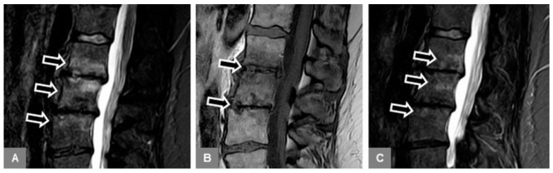
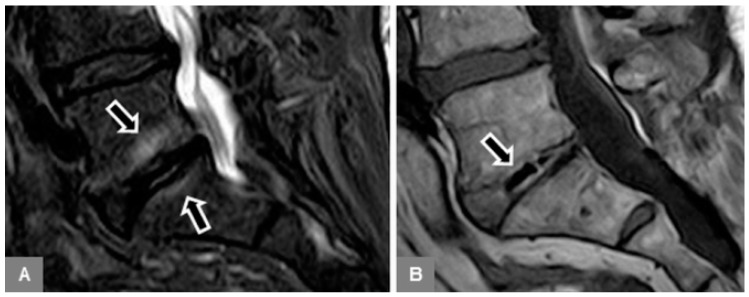
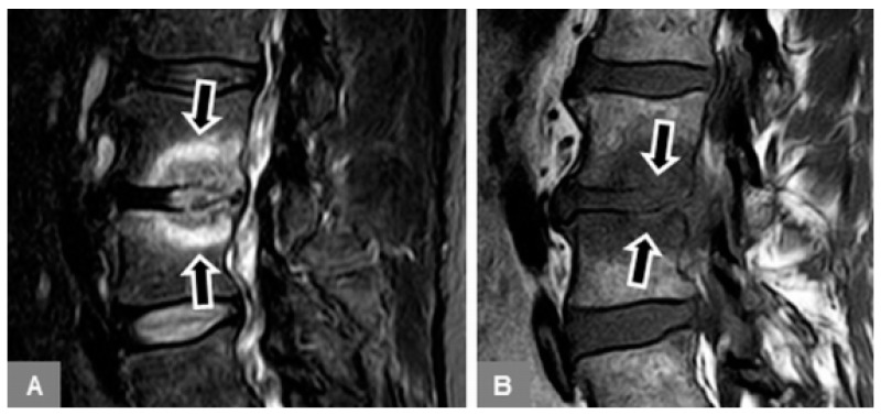
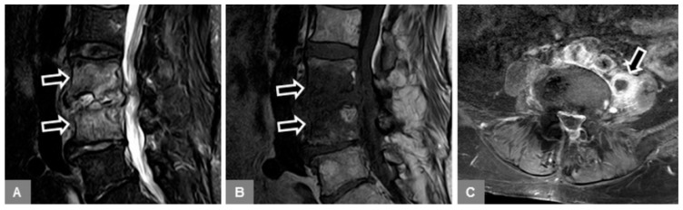
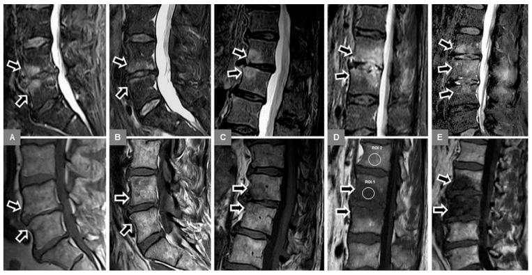
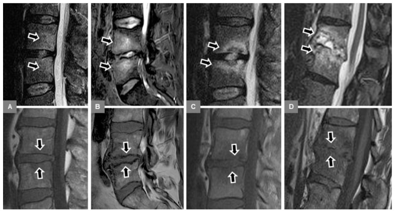
On magnetic resonance (MR) imaging, Modic type 1 (MT1) endplate changes and infectious spondylodiscitis share similar findings. Therefore, this study investigated vertebral bone marrow and endplate changes to enable their differentiation. The lumbar spine MR examinations of 91 adult patients were retrospectively included: 39 with MT1; 19 with early spondylodiscitis without abscess; and 33 with advanced spondylodiscitis with abscess. The assessment included percentage of bone marrow edema on sagittal short tau inversion recovery images, and the signal ratio of edema to unaffected bone and endplate contour (normal; irregular, yet intact; blurred; destructive) on sagittal unenhanced T1-weighted images. Differences were tested for statistical significance by Chi-square test and mixed model analysis of variance. The MR diagnostic accuracy in differentiating MT1 and spondylodiscitis was assessed by cross-tabulation and receiver-operating characteristic analysis. The endplate contours, edema extents, and T1-signal ratios of MT1 (extent, 31.96%; ratio, 0.83) were significantly different ( < 0.001) from early spondylodiscitis (56.42%; 0.60), and advanced spondylodiscitis (91.84%; 0.61). The highest diagnostic accuracy (sensitivity, 94.87%; specificity, 94.23%; accuracy, 94.51%) in identifying MT1 was provided by an irregular, yet intact endplate contour. This may be a useful MR feature for the differentiation between MT1 and spondylodiscitis, particularly in its early stage.

摘要

在磁共振成像(MR)中,Modic 1型(MT1)终板改变与感染性脊椎椎间盘炎有相似表现。因此,本研究对椎体骨髓和终板改变进行了调查,以便对它们进行鉴别。回顾性纳入了91例成年患者的腰椎MR检查:39例为MT1;19例为早期无脓肿的脊椎椎间盘炎;33例为晚期有脓肿的脊椎椎间盘炎。评估包括矢状面短Tau反转恢复图像上骨髓水肿的百分比,以及矢状面未增强T1加权图像上水肿与未受影响的骨骼和终板轮廓(正常;不规则但完整;模糊;破坏)的信号比。通过卡方检验和方差混合模型分析检验差异的统计学意义。通过交叉表和受试者操作特征分析评估MR鉴别MT1和脊椎椎间盘炎的诊断准确性。MT1的终板轮廓、水肿范围和T1信号比(范围,31.96%;比率,0.83)与早期脊椎椎间盘炎(56.42%;0.60)和晚期脊椎椎间盘炎(91.84%;0.61)有显著差异(<0.001)。识别MT1的最高诊断准确性(敏感性,94.87%;特异性,94.23%;准确性,94.51%)由不规则但完整的终板轮廓提供。这可能是鉴别MT1和脊椎椎间盘炎的一个有用的MR特征,特别是在早期阶段。

https://cdn.ncbi.nlm.nih.gov/pmc/blobs/b700/7141365/42317d95d266/jcm-09-00826-g006.jpg
https://cdn.ncbi.nlm.nih.gov/pmc/blobs/b700/7141365/e3affb3fb067/jcm-09-00826-g001.jpg
https://cdn.ncbi.nlm.nih.gov/pmc/blobs/b700/7141365/d92eccdae660/jcm-09-00826-g002.jpg
https://cdn.ncbi.nlm.nih.gov/pmc/blobs/b700/7141365/668bed5b393e/jcm-09-00826-g003.jpg
https://cdn.ncbi.nlm.nih.gov/pmc/blobs/b700/7141365/9b4957f0ebab/jcm-09-00826-g004.jpg
https://cdn.ncbi.nlm.nih.gov/pmc/blobs/b700/7141365/ec2982d8d3b0/jcm-09-00826-g005.jpg
https://cdn.ncbi.nlm.nih.gov/pmc/blobs/b700/7141365/42317d95d266/jcm-09-00826-g006.jpg

相似文献

1
Vertebral Bone Marrow and Endplate Assessment on MR Imaging for the Differentiation of Modic Type 1 Endplate Changes and Infectious Spondylodiscitis.磁共振成像对椎体骨髓和终板的评估用于鉴别Modic 1型终板改变与感染性脊椎椎间盘炎
J Clin Med. 2020 Mar 18;9(3):826. doi: 10.3390/jcm9030826.
2
Reliability of diffusion weighted MR imaging in differentiating degenerative and infectious end plate changes.弥散加权磁共振成像在鉴别退行性和感染性终板改变中的可靠性。
Radiol Oncol. 2010 Jun;44(2):97-102. doi: 10.2478/v10019-010-0006-z. Epub 2010 May 24.
3
Paraspinal soft tissue edema ratio: An accurate marker for early lumbar spine spondylodiscitis on an unenhanced MRI.椎旁软组织水肿率:未增强磁共振成像上早期腰椎椎体骨髓炎的准确标志物。
Clin Imaging. 2022 Jun;86:38-42. doi: 10.1016/j.clinimag.2022.03.009. Epub 2022 Mar 14.
4
Proton Density Fat Fraction Spine MRI for Differentiation of Erosive Vertebral Endplate Degeneration and Infectious Spondylitis.用于鉴别侵蚀性椎体终板退变和感染性脊柱炎的质子密度脂肪分数脊柱磁共振成像
Diagnostics (Basel). 2021 Dec 30;12(1):78. doi: 10.3390/diagnostics12010078.
5
FDG positron emission tomography for differentiation of degenerative and infectious endplate abnormalities in the lumbar spine detected on MR imaging.利用氟代脱氧葡萄糖正电子发射断层扫描鉴别腰椎磁共振成像检测到的退行性和感染性终板异常。
AJR Am J Roentgenol. 2002 Nov;179(5):1151-7. doi: 10.2214/ajr.179.5.1791151.
6
Modic changes in vertebral endplates: a comparison of MR imaging and multislice CT.椎体终板的Modic改变:磁共振成像与多层螺旋CT的比较
Skeletal Radiol. 2009 Feb;38(2):141-7. doi: 10.1007/s00256-008-0590-9. Epub 2008 Sep 20.
7
Magnetic Resonance Imaging in Differentatial Diagnosis of Pyogenic Spondylodiscitis and Tuberculous Spondylodiscitis.磁共振成像在化脓性脊椎椎间盘炎与结核性脊椎椎间盘炎鉴别诊断中的应用
Pol J Radiol. 2017 Feb 10;82:71-87. doi: 10.12659/PJR.899606. eCollection 2017.
8
Effects of sagittal endplate shape on lumbar segmental mobility as evaluated by kinetic magnetic resonance imaging.通过动态磁共振成像评估矢状面终板形状对腰椎节段活动度的影响。
Spine (Phila Pa 1976). 2014 Aug 1;39(17):E1035-41. doi: 10.1097/BRS.0000000000000419.
9
Reactive endplate marrow changes: a systematic morphologic and epidemiologic evaluation.反应性终板骨髓改变:一项系统的形态学和流行病学评估
Skeletal Radiol. 2005 Mar;34(3):125-9. doi: 10.1007/s00256-004-0886-3. Epub 2005 Jan 13.
10
Diffusion magnetic resonance imaging to differentiate degenerative from infectious endplate abnormalities in the lumbar spine.扩散磁共振成像区分腰椎退行性与感染性终板异常。
Spine (Phila Pa 1976). 2011 Feb 1;36(3):E198-202. doi: 10.1097/BRS.0b013e3181d5ff05.

引用本文的文献

1
Diagnostic imaging confusion in infectious spondylitis.感染性脊柱炎的诊断性影像学混淆
J Orthop Surg Res. 2025 May 21;20(1):496. doi: 10.1186/s13018-025-05781-5.
2
Sequestered Lumbar Disc Mimicking Psoas Abscess: A Case Report.隐匿性腰椎间盘突出症酷似腰大肌脓肿:一例报告
Cureus. 2025 Feb 27;17(2):e79781. doi: 10.7759/cureus.79781. eCollection 2025 Feb.
3
Musculoskeletal Infection: The Great Mimickers on Imaging.肌肉骨骼感染:影像学上的“模仿大师”

本文引用的文献

1
Impact of MRI, CT, and Clinical Characteristics on Microbial Pathogen Detection Using CT-Guided Biopsy for Suspected Spondylodiscitis.磁共振成像(MRI)、计算机断层扫描(CT)及临床特征对采用CT引导下活检诊断疑似脊椎椎间盘炎时微生物病原体检测的影响
J Clin Med. 2019 Dec 21;9(1):32. doi: 10.3390/jcm9010032.
2
Infectious Spondylitis Mimics: Mechanisms of Disease and Imaging Findings.感染性脊柱炎的模仿者:发病机制与影像学表现
Semin Ultrasound CT MR. 2018 Dec;39(6):587-604. doi: 10.1053/j.sult.2018.11.006. Epub 2018 Nov 15.
3
Imaging of Spondylodiscitis.脊柱骨髓炎的影像学表现。
J Clin Med. 2024 Sep 13;13(18):5424. doi: 10.3390/jcm13185424.
4
From Modic to Disc Endplate Bone Marrow Complex - The Natural Course and Clinical Implication of Vertebral Endplate Changes.从莫迪克改变到椎间盘终板骨髓复合体——椎体终板改变的自然病程及临床意义
Global Spine J. 2025 Jan;15(1):196-209. doi: 10.1177/21925682241271440. Epub 2024 Aug 1.
5
Can Hematological Inflammatory Indices Be Used to Differentiate Modic Type 1 Changes from Brucella Spondylodiscitis?能否通过血液学炎症指标区分 Modic 型 1 改变与布鲁氏菌性脊椎炎?
Medicina (Kaunas). 2024 Jul 14;60(7):1131. doi: 10.3390/medicina60071131.
6
Imaging assessment of spine infection.脊柱感染的影像学评估。
Skeletal Radiol. 2024 Oct;53(10):2067-2079. doi: 10.1007/s00256-023-04558-3. Epub 2024 Jan 16.
7
Study of the Distribution of Lumbar Modic Changes in Patients with Low Back Pain and Correlation with Lumbar Degeneration Diseases.腰痛患者腰椎Modic改变的分布及其与腰椎退变疾病相关性的研究
J Pain Res. 2023 Nov 6;16:3725-3733. doi: 10.2147/JPR.S430792. eCollection 2023.
8
New Concepts in Diagnosis, Risk Factors and Work Ability in Patients with Hematogenous Spinal Infection.血源性脊柱感染患者诊断、危险因素及工作能力的新概念
J Clin Med. 2022 Sep 17;11(18):5470. doi: 10.3390/jcm11185470.
9
Imaging and Clinical Parameters for Distinction between Infected and Non-Infected Fluid Collections in CT: Prospective Study Using Extended Microbiological Approach.CT 中区分感染性与非感染性液体积聚的影像学和临床参数:采用扩展微生物学方法的前瞻性研究
Diagnostics (Basel). 2022 Feb 14;12(2):493. doi: 10.3390/diagnostics12020493.
10
Proton Density Fat Fraction Spine MRI for Differentiation of Erosive Vertebral Endplate Degeneration and Infectious Spondylitis.用于鉴别侵蚀性椎体终板退变和感染性脊柱炎的质子密度脂肪分数脊柱磁共振成像
Diagnostics (Basel). 2021 Dec 30;12(1):78. doi: 10.3390/diagnostics12010078.
Semin Nucl Med. 2018 Mar;48(2):131-147. doi: 10.1053/j.semnuclmed.2017.11.001. Epub 2018 Feb 6.
4
Effect of Zoledronic Acid and Denosumab in Patients With Low Back Pain and Modic Change: A Proof-of-Principle Trial.唑来膦酸和地舒单抗治疗腰痛伴 Modic 改变患者的效果:一项原理验证试验。
J Bone Miner Res. 2018 May;33(5):773-782. doi: 10.1002/jbmr.3376. Epub 2018 Feb 2.
5
Modic type 1 change is an autoimmune response that requires a proinflammatory milieu provided by the 'Modic disc'.Modic 型 1 改变是一种自身免疫反应,需要由“Modic 盘”提供的促炎环境。
Spine J. 2018 May;18(5):831-844. doi: 10.1016/j.spinee.2017.12.004. Epub 2017 Dec 15.
6
Modic Type 1 Changes: Detection Performance of Fat-Suppressed Fluid-Sensitive MRI Sequences.Modic 1型改变:脂肪抑制液体敏感型MRI序列的检测性能
Rofo. 2018 Feb;190(2):152-160. doi: 10.1055/s-0043-118130. Epub 2017 Nov 27.
7
Inflammatory response of disc cells against Propionibacterium acnes depends on the presence of lumbar Modic changes.椎间盘细胞对痤疮丙酸杆菌的炎症反应取决于腰椎Modic改变的存在情况。
Eur Spine J. 2018 May;27(5):1013-1020. doi: 10.1007/s00586-017-5291-4. Epub 2017 Sep 7.
8
Gadolinium deposition in the brain: summary of evidence and recommendations.脑内钆沉积:证据总结和建议。
Lancet Neurol. 2017 Jul;16(7):564-570. doi: 10.1016/S1474-4422(17)30158-8. Epub 2017 Jun 13.
9
Magnetic resonance imaging of bacterial and tuberculous spondylodiscitis with associated complications and non-infectious spinal pathology mimicking infections: a pictorial review.细菌性和结核性脊椎椎间盘炎伴相关并发症及类似感染的非感染性脊柱病变的磁共振成像:图文综述
BMC Musculoskelet Disord. 2017 Jun 5;18(1):244. doi: 10.1186/s12891-017-1608-z.
10
Modic Type 1 Vertebral Endplate Changes: Injury, Inflammation, or Infection?Modic 型 1 椎体终板改变:损伤、炎症还是感染?
AJR Am J Roentgenol. 2017 Jul;209(1):167-170. doi: 10.2214/AJR.16.17403. Epub 2017 Apr 12.