Laboratory of Immunology and Infectious Diseases, Graduate School of Medical Science and Engineering, KAIST, Daejeon, Republic of Korea.
PLoS One. 2012;7(8):e43960. doi: 10.1371/journal.pone.0043960. Epub 2012 Aug 24.
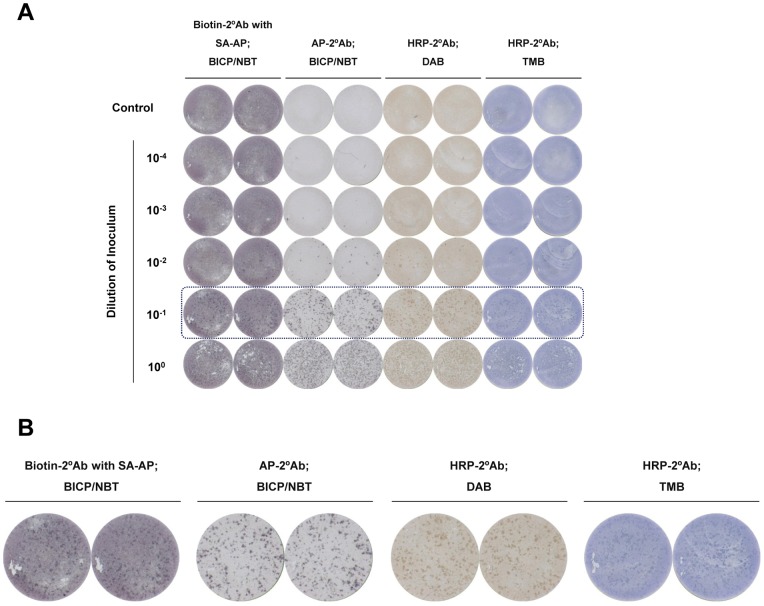
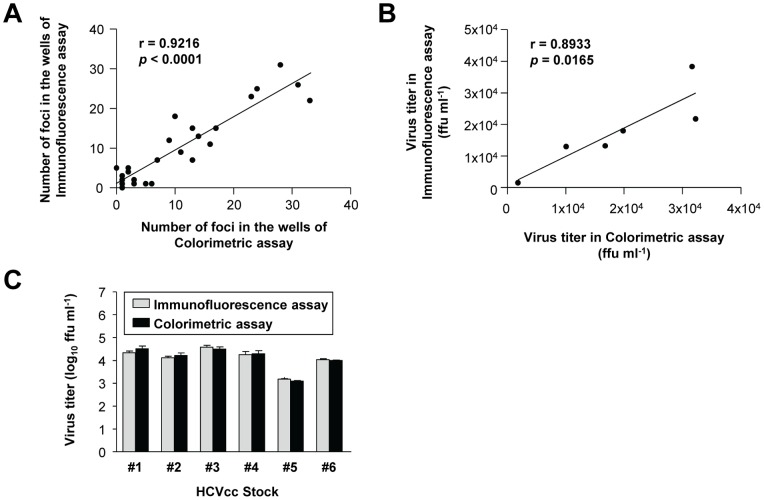
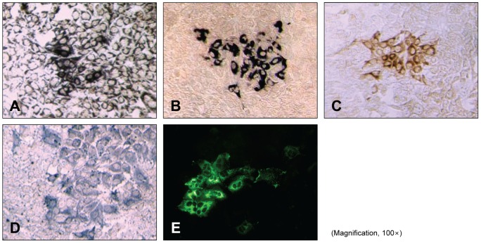
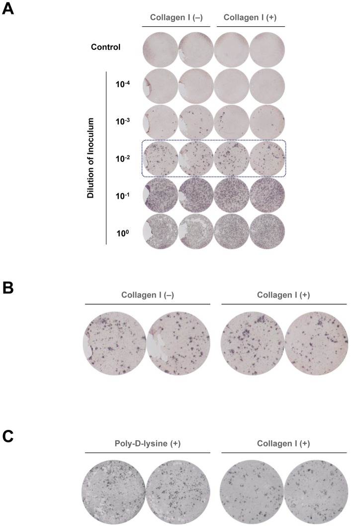
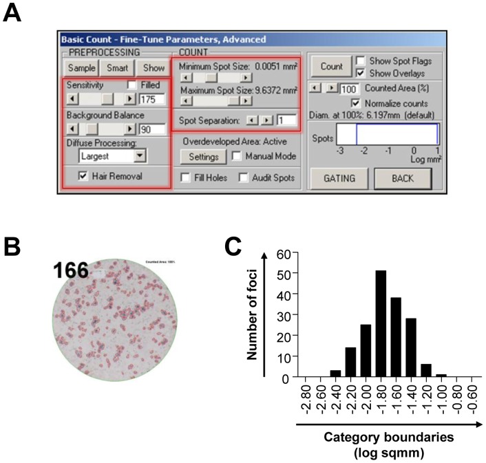
Hepatitis C virus (HCV) infection is the leading cause of liver transplantation in Western countries. Studies of HCV infection using cell culture-produced HCV (HCVcc) in vitro systems require quantification of infectious HCV virions, which has conventionally been performed by immunofluorescence-based focus-forming assay with manual foci counting; however, this is a laborious and time-consuming procedure with potentially biased results. In the present study, we established and optimized a method for convenient and objective quantification of HCV virions by colorimetric focus-forming assay with automated focus counting by image analysis. In testing different enzymes and chromogenic substrates, we obtained superior foci development using alkaline phosphatase-conjugated secondary antibody with BCIP/NBT chromogenic substrate. We additionally found that type I collagen coating minimized cell detachment during vigorous washing of the assay plate. After the colorimetric focus-forming assay, the foci number was determined using an ELISpot reader and image analysis software. The foci number and the calculated viral titer determined by this method strongly correlated with those determined by immunofluorescence-based focus-forming assay and manual foci counting. These results indicate that colorimetric focus-forming assay with automated focus counting by image analysis is applicable as a more-efficient and objective method for quantification of infectious HCV virions.
丙型肝炎病毒 (HCV) 感染是西方国家肝移植的主要原因。使用细胞培养产生的丙型肝炎病毒 (HCVcc) 在体外系统研究 HCV 感染需要定量感染性 HCV 病毒颗粒,这通常通过基于免疫荧光的焦点形成测定法和手动焦点计数来完成;然而,这是一项繁琐且耗时的过程,结果可能存在偏差。在本研究中,我们建立并优化了一种通过比色焦点形成测定法和自动焦点计数的图像分析进行 HCV 病毒颗粒的定量的方法。在测试不同的酶和显色底物时,我们使用碱性磷酸酶缀合的二级抗体和 BCIP/NBT 显色底物获得了更好的焦点发展。我们还发现,I 型胶原涂层可最大程度地减少测定板剧烈洗涤过程中的细胞脱落。比色焦点形成测定法后,使用 ELISpot 读取器和图像分析软件确定焦点数量。该方法确定的焦点数量和计算的病毒滴度与基于免疫荧光的焦点形成测定法和手动焦点计数确定的焦点数量和计算的病毒滴度密切相关。这些结果表明,通过图像分析进行自动焦点计数的比色焦点形成测定法可作为一种更有效的、客观的定量传染性 HCV 病毒颗粒的方法。