• 文献检索
  • 文档翻译
  • 深度研究
  • 学术资讯
  • Suppr Zotero 插件Zotero 插件
  • 邀请有礼
  • 套餐&价格
  • 历史记录
应用&插件
Suppr Zotero 插件Zotero 插件浏览器插件Mac 客户端Windows 客户端微信小程序
定价
高级版会员购买积分包购买API积分包
服务
文献检索文档翻译深度研究API 文档MCP 服务
关于我们
关于 Suppr公司介绍联系我们用户协议隐私条款
关注我们

Suppr 超能文献

核心技术专利:CN118964589B侵权必究
粤ICP备2023148730 号-1Suppr @ 2026

文献检索

告别复杂PubMed语法,用中文像聊天一样搜索,搜遍4000万医学文献。AI智能推荐,让科研检索更轻松。

立即免费搜索

文件翻译

保留排版,准确专业,支持PDF/Word/PPT等文件格式,支持 12+语言互译。

免费翻译文档

深度研究

AI帮你快速写综述,25分钟生成高质量综述,智能提取关键信息,辅助科研写作。

立即免费体验

基于强度匹配和完全异常值去除的胎儿脑 MRI 重建。

Reconstruction of fetal brain MRI with intensity matching and complete outlier removal.

机构信息

Institute of Biomedical Engineering, Department of Engineering Science, University of Oxford, UK.

出版信息

Med Image Anal. 2012 Dec;16(8):1550-64. doi: 10.1016/j.media.2012.07.004. Epub 2012 Aug 9.

DOI:10.1016/j.media.2012.07.004
PMID:22939612
原文链接:https://pmc.ncbi.nlm.nih.gov/articles/PMC4067058/
Abstract

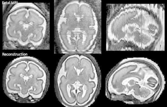
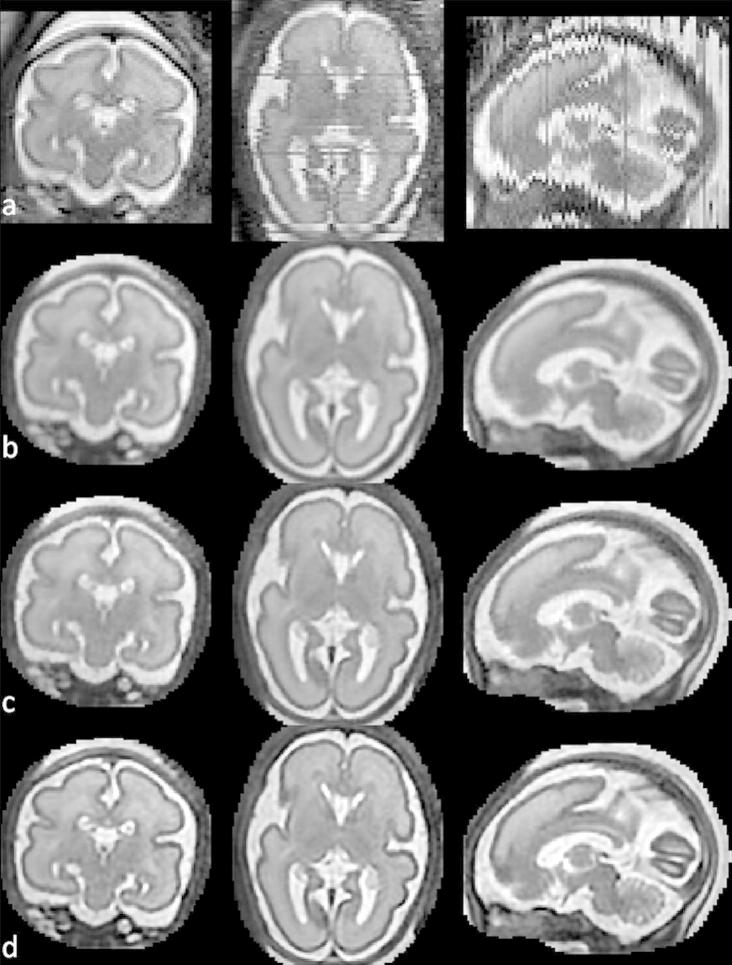
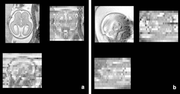
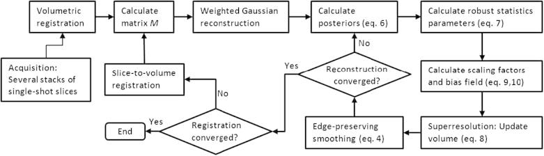
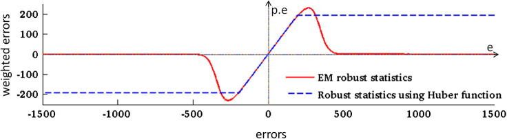
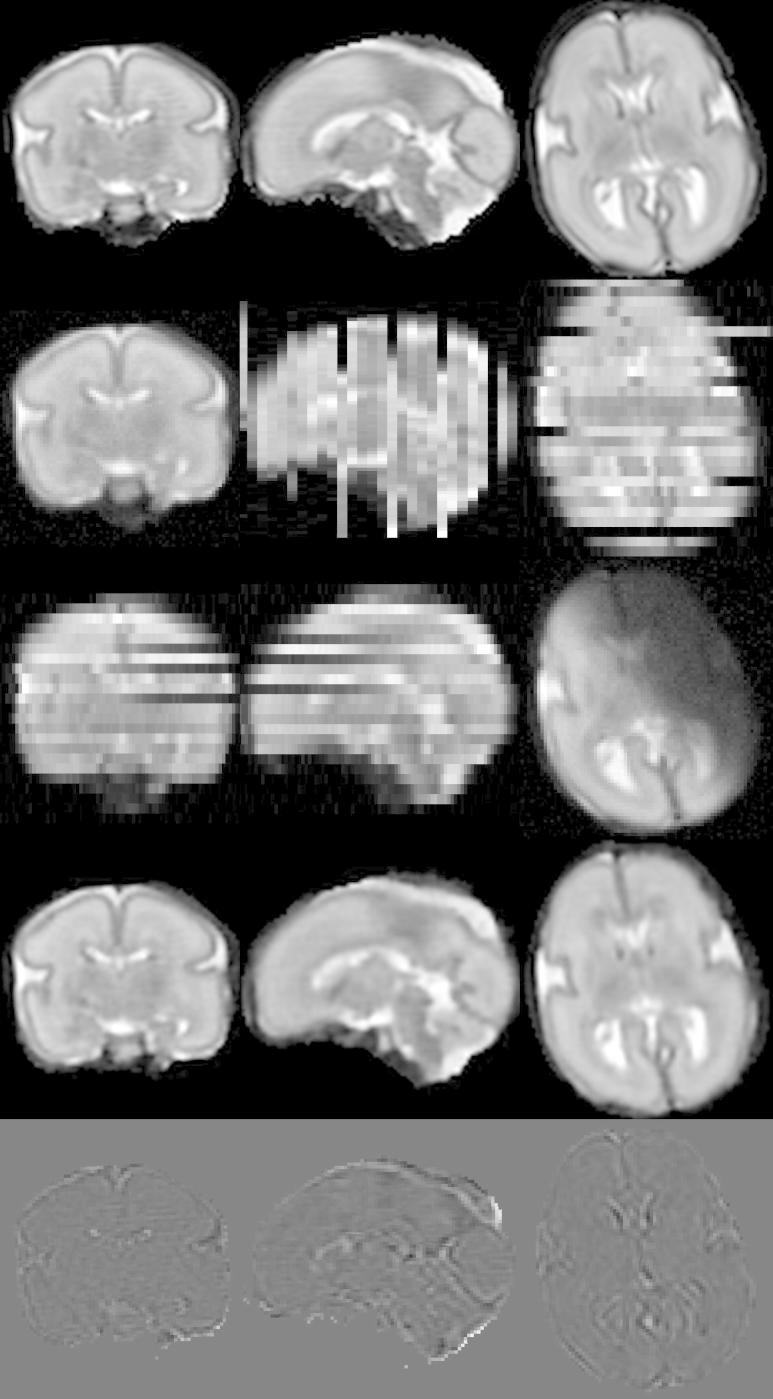
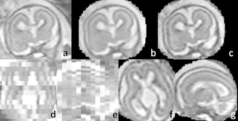
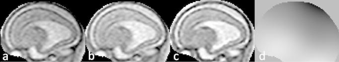
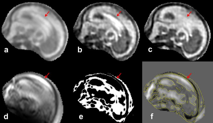
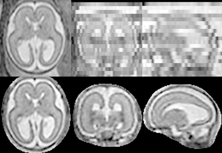
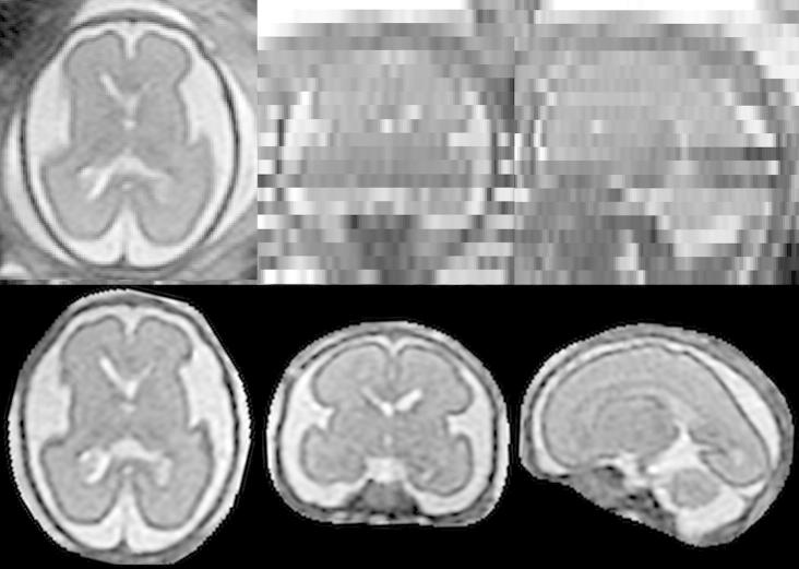
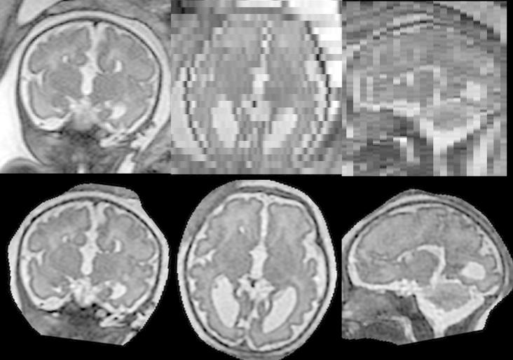
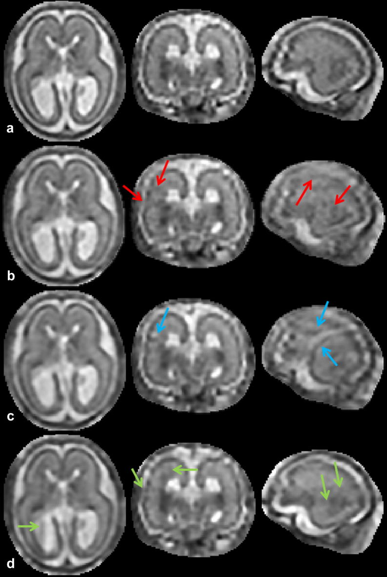
We propose a method for the reconstruction of volumetric fetal MRI from 2D slices, comprising super-resolution reconstruction of the volume interleaved with slice-to-volume registration to correct for the motion. The method incorporates novel intensity matching of acquired 2D slices and robust statistics which completely excludes identified misregistered or corrupted voxels and slices. The reconstruction method is applied to motion-corrupted data simulated from MRI of a preterm neonate, as well as 10 clinically acquired thick-slice fetal MRI scans and three scan-sequence optimized thin-slice fetal datasets. The proposed method produced high quality reconstruction results from all the datasets to which it was applied. Quantitative analysis performed on simulated and clinical data shows that both intensity matching and robust statistics result in statistically significant improvement of super-resolution reconstruction. The proposed novel EM-based robust statistics also improves the reconstruction when compared to previously proposed Huber robust statistics. The best results are obtained when thin-slice data and the correct approximation of the point spread function is used. This paper addresses the need for a comprehensive reconstruction algorithm of 3D fetal MRI, so far lacking in the scientific literature.

摘要

我们提出了一种从 2D 切片重建胎儿 MRI 体数据集的方法,该方法包括体积超分辨率重建和用于纠正运动的层间体积到层间的配准。该方法采用新颖的采集二维切片的强度匹配和稳健统计学方法,完全排除了已识别的配准错误或损坏的体素和切片。该重建方法应用于从早产儿 MRI 模拟的运动伪影数据,以及 10 个临床采集的厚层胎儿 MRI 扫描和三个扫描序列优化的薄层胎儿数据集。所提出的方法对所有应用数据集都产生了高质量的重建结果。对模拟和临床数据的定量分析表明,强度匹配和稳健统计学都导致超分辨率重建的显著改善。与以前提出的 Huber 稳健统计学相比,所提出的基于 EM 的稳健统计学也提高了重建效果。当使用薄层数据和正确的点扩散函数近似值时,可获得最佳结果。本文针对目前科学文献中缺乏全面的 3D 胎儿 MRI 重建算法的问题,提出了一种新的解决方案。

https://cdn.ncbi.nlm.nih.gov/pmc/blobs/f246/4067058/c043ac1a7c12/gr13.jpg
https://cdn.ncbi.nlm.nih.gov/pmc/blobs/f246/4067058/1f6f8a90afd7/fx1.jpg
https://cdn.ncbi.nlm.nih.gov/pmc/blobs/f246/4067058/7aa88ec43ed2/gr1.jpg
https://cdn.ncbi.nlm.nih.gov/pmc/blobs/f246/4067058/a76e24b30432/gr2.jpg
https://cdn.ncbi.nlm.nih.gov/pmc/blobs/f246/4067058/11f205ccd60a/gr3.jpg
https://cdn.ncbi.nlm.nih.gov/pmc/blobs/f246/4067058/9a98f0255067/gr4.jpg
https://cdn.ncbi.nlm.nih.gov/pmc/blobs/f246/4067058/7e18a3c83dfd/gr5.jpg
https://cdn.ncbi.nlm.nih.gov/pmc/blobs/f246/4067058/76bc41422c72/gr6.jpg
https://cdn.ncbi.nlm.nih.gov/pmc/blobs/f246/4067058/2a9fdc91873c/gr7.jpg
https://cdn.ncbi.nlm.nih.gov/pmc/blobs/f246/4067058/3a38a00fedfb/gr8.jpg
https://cdn.ncbi.nlm.nih.gov/pmc/blobs/f246/4067058/30fed754c1b1/gr9.jpg
https://cdn.ncbi.nlm.nih.gov/pmc/blobs/f246/4067058/1255583f25d2/gr10.jpg
https://cdn.ncbi.nlm.nih.gov/pmc/blobs/f246/4067058/cd310b103c48/gr11.jpg
https://cdn.ncbi.nlm.nih.gov/pmc/blobs/f246/4067058/5a719ed7df02/gr12.jpg
https://cdn.ncbi.nlm.nih.gov/pmc/blobs/f246/4067058/c043ac1a7c12/gr13.jpg

相似文献

1
Reconstruction of fetal brain MRI with intensity matching and complete outlier removal.基于强度匹配和完全异常值去除的胎儿脑 MRI 重建。
Med Image Anal. 2012 Dec;16(8):1550-64. doi: 10.1016/j.media.2012.07.004. Epub 2012 Aug 9.
2
Intersection based motion correction of multislice MRI for 3-D in utero fetal brain image formation.基于交叠的多层 MRI 运动校正用于三维宫内胎儿脑图像形成。
IEEE Trans Med Imaging. 2010 Jan;29(1):146-58. doi: 10.1109/TMI.2009.2030679. Epub 2009 Sep 9.
3
Automated template-based brain localization and extraction for fetal brain MRI reconstruction.基于模板的自动化胎儿脑磁共振成像重建的脑定位与提取
Neuroimage. 2017 Jul 15;155:460-472. doi: 10.1016/j.neuroimage.2017.04.004. Epub 2017 Apr 11.
4
NeSVoR: Implicit Neural Representation for Slice-to-Volume Reconstruction in MRI.NeSVoR:MRI 中用于切片到体素重建的隐式神经表示。
IEEE Trans Med Imaging. 2023 Jun;42(6):1707-1719. doi: 10.1109/TMI.2023.3236216. Epub 2023 Jun 1.
5
Automated fetal brain segmentation from 2D MRI slices for motion correction.基于 2D MRI 切片的自动胎儿脑分割用于运动校正。
Neuroimage. 2014 Nov 1;101:633-43. doi: 10.1016/j.neuroimage.2014.07.023. Epub 2014 Jul 22.
6
AFFIRM: Affinity Fusion-Based Framework for Iteratively Random Motion Correction of Multi-Slice Fetal Brain MRI.AFFIRM:基于亲和融合的多切片胎儿脑磁共振成像迭代随机运动校正框架。
IEEE Trans Med Imaging. 2023 Jan;42(1):209-219. doi: 10.1109/TMI.2022.3208277. Epub 2022 Dec 29.
7
Automated 3D reconstruction of the fetal thorax in the standard atlas space from motion-corrupted MRI stacks for 21-36 weeks GA range.从 21 周到 36 周 GA 范围内运动伪影的 MRI 堆栈中自动在标准图谱空间重建胎儿胸腔的 3D 结构。
Med Image Anal. 2022 Aug;80:102484. doi: 10.1016/j.media.2022.102484. Epub 2022 May 25.
8
Fetal brain volumetry through MRI volumetric reconstruction and segmentation.MRI 容积重建和分割技术测量胎儿脑容量。
Int J Comput Assist Radiol Surg. 2011 May;6(3):329-39. doi: 10.1007/s11548-010-0512-x. Epub 2010 Jul 13.
9
A novel approach to high resolution fetal brain MR imaging.一种用于高分辨率胎儿脑部磁共振成像的新方法。
Med Image Comput Comput Assist Interv. 2005;8(Pt 1):548-55. doi: 10.1007/11566465_68.
10
Three-dimensional visualisation of the fetal heart using prenatal MRI with motion-corrected slice-volume registration: a prospective, single-centre cohort study.使用带运动校正的层面容积配准的产前 MRI 对胎儿心脏进行三维可视化:一项前瞻性、单中心队列研究。
Lancet. 2019 Apr 20;393(10181):1619-1627. doi: 10.1016/S0140-6736(18)32490-5. Epub 2019 Mar 22.

引用本文的文献

1
An MRI Atlas of the Human Fetal Brain: Reference and Segmentation Tools for Fetal Brain MRI Analysis.《人类胎儿脑部磁共振成像图谱:胎儿脑部磁共振成像分析的参考与分割工具》
ArXiv. 2025 Aug 28:arXiv:2508.15034v2.
2
Mechanics of the Spatiotemporal Evolution of Sulcal Pits in the Folding Brain.折叠大脑中脑沟凹陷的时空演化机制
Hum Brain Mapp. 2025 Sep;46(13):e70332. doi: 10.1002/hbm.70332.
3
Perinatal development of structural thalamocortical connectivity.丘脑皮质结构连接的围产期发育

本文引用的文献

1
Spatiotemporal morphometry of adjacent tissue layers with application to the study of sulcal formation.相邻组织层的时空形态测量及其在脑沟形成研究中的应用
Med Image Comput Comput Assist Interv. 2011;14(Pt 2):476-83. doi: 10.1007/978-3-642-23629-7_58.
2
Early folding patterns and asymmetries of the normal human brain detected from in utero MRI.从宫内 MRI 检测到的正常人类大脑的早期折叠模式和不对称性。
Cereb Cortex. 2012 Jan;22(1):13-25. doi: 10.1093/cercor/bhr053. Epub 2011 May 12.
3
Mapping fetal brain development in utero using magnetic resonance imaging: the Big Bang of brain mapping.
Imaging Neurosci (Camb). 2025 Jan 8;3. doi: 10.1162/imag_a_00418. eCollection 2025.
4
HAITCH: A framework for distortion and motion correction in fetal multi-shell diffusion-weighted MRI.HAITCH:胎儿多壳扩散加权磁共振成像中畸变与运动校正的框架
Imaging Neurosci (Camb). 2025 Feb 26;3. doi: 10.1162/imag_a_00490. eCollection 2025.
5
Biometry and volumetry in multi-centric fetal brain magnetic resonance imaging: assessing the bias of super-resolution reconstruction.多中心胎儿脑磁共振成像中的生物测量与容积测量:评估超分辨率重建的偏差
Pediatr Radiol. 2025 Aug 8. doi: 10.1007/s00247-025-06347-7.
6
Cord blood levels of insulin-like growth factor-1 and insulin-like growth factor binding protein-3 correlate with perinatal brain development in fetal congenital heart disease.胎儿先天性心脏病中,脐血胰岛素样生长因子-1和胰岛素样生长因子结合蛋白-3水平与围产期脑发育相关。
Ultrasound Obstet Gynecol. 2025 Aug;66(2):200-209. doi: 10.1002/uog.29271. Epub 2025 Jun 28.
7
Common genetic variation associated with adult subcortical brain volume is also associated with subcortical brain volume at birth.与成人皮质下脑容量相关的常见基因变异也与出生时的皮质下脑容量有关。
Front Neurosci. 2025 Jun 10;19:1546845. doi: 10.3389/fnins.2025.1546845. eCollection 2025.
8
Placental histology, perioperative brain development, and neurodevelopmental outcome at 1 year of age in patients undergoing neonatal cardiac surgery-is there an association?新生儿心脏手术患者的胎盘组织学、围手术期脑发育及1岁时的神经发育结局——它们之间有关联吗?
Front Cardiovasc Med. 2025 May 30;12:1556289. doi: 10.3389/fcvm.2025.1556289. eCollection 2025.
9
Differential patterns of cortical expansion in fetal and preterm brain development.胎儿和早产儿大脑发育过程中皮质扩张的差异模式。
bioRxiv. 2025 May 21:2025.05.19.653958. doi: 10.1101/2025.05.19.653958.
10
Early life stress impairs hippocampal subfield myelination.早年生活应激会损害海马亚区的髓鞘形成。
Commun Biol. 2025 May 22;8(1):785. doi: 10.1038/s42003-025-08165-x.
利用磁共振成像对胎儿大脑发育进行映射:大脑映射的大爆炸。
Annu Rev Biomed Eng. 2011 Aug 15;13:345-68. doi: 10.1146/annurev-bioeng-071910-124654.
4
Quantitative in vivo MRI measurement of cortical development in the fetus.胎儿皮质发育的活体定量 MRI 测量。
Brain Struct Funct. 2012 Jan;217(1):127-39. doi: 10.1007/s00429-011-0325-x. Epub 2011 May 12.
5
Bias field inconsistency correction of motion-scattered multislice MRI for improved 3D image reconstruction.运动散射多切片 MRI 的偏置场不一致性校正,以改善 3D 图像重建。
IEEE Trans Med Imaging. 2011 Sep;30(9):1704-12. doi: 10.1109/TMI.2011.2143724. Epub 2011 Apr 19.
6
Local tissue growth patterns underlying normal fetal human brain gyrification quantified in utero.在子宫内定量研究正常胎儿人类大脑脑回形成的局部组织生长模式。
J Neurosci. 2011 Feb 23;31(8):2878-87. doi: 10.1523/JNEUROSCI.5458-10.2011.
7
On super-resolution for fetal brain MRI.关于胎儿脑磁共振成像的超分辨率
Med Image Comput Comput Assist Interv. 2010;13(Pt 2):355-62. doi: 10.1007/978-3-642-15745-5_44.
8
Fetal brain volumetry through MRI volumetric reconstruction and segmentation.MRI 容积重建和分割技术测量胎儿脑容量。
Int J Comput Assist Radiol Surg. 2011 May;6(3):329-39. doi: 10.1007/s11548-010-0512-x. Epub 2010 Jul 13.
9
Robust super-resolution volume reconstruction from slice acquisitions: application to fetal brain MRI.从切片采集进行稳健的超分辨率体积重建:在胎儿脑 MRI 中的应用。
IEEE Trans Med Imaging. 2010 Oct;29(10):1739-58. doi: 10.1109/TMI.2010.2051680. Epub 2010 Jun 7.
10
Intersection based motion correction of multislice MRI for 3-D in utero fetal brain image formation.基于交叠的多层 MRI 运动校正用于三维宫内胎儿脑图像形成。
IEEE Trans Med Imaging. 2010 Jan;29(1):146-58. doi: 10.1109/TMI.2009.2030679. Epub 2009 Sep 9.