Division of Cellular Biosignal Sciences, Department of Biochemistry, Iwate Medical University, 2-1-1 Nishitokuta, Yahaba-cho, Shiwa-gun, Iwate 028-3694, Japan.
Int J Biol Sci. 2012;8(7):1062-74. doi: 10.7150/ijbs.4488. Epub 2012 Aug 22.
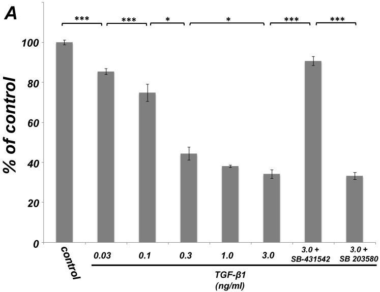
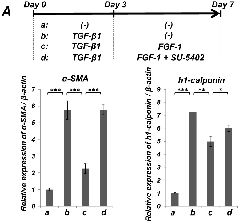
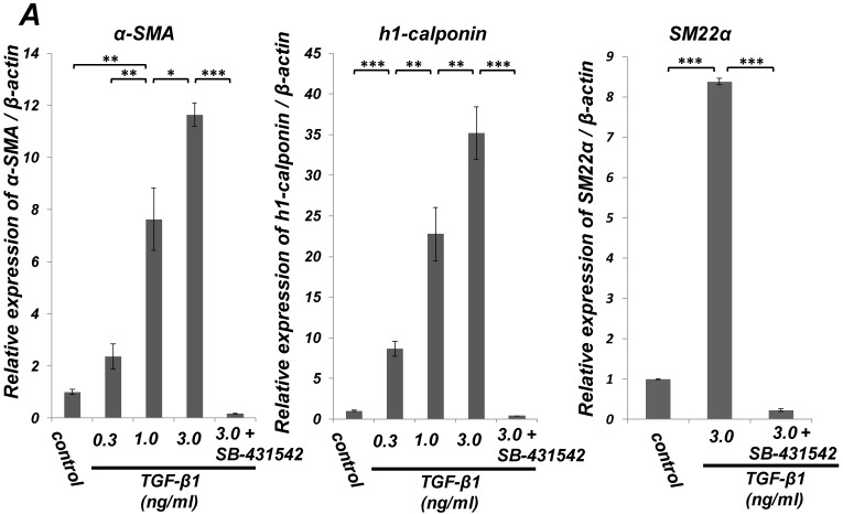
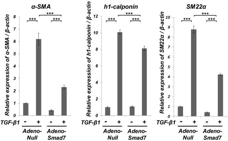
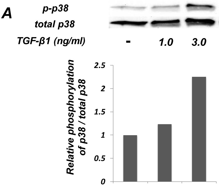
The periodontal ligament (PDL) is a fibrous connective tissue that attaches the tooth to the alveolar bone. We previously demonstrated the ability of PDL fibroblast-like cells to construct an endothelial cell (EC) marker-positive blood vessel-like structure, indicating the potential of fibroblastic lineage cells in PDL tissue as precursors of endothelial progenitor cells (EPCs) to facilitate the construction of a vascular system around damaged PDL tissue. A vascular regeneration around PDL tissue needs proliferation of vascular progenitor cells and the subsequent differentiation of the cells. Transforming growth factor-β (TGF-β) is known as an inducer of endothelial-mesenchymal transition (EndMT), however, it remains to be clarified what kinds of TGF-β signals affect growth and mesenchymal differentiation of PDL-derived EPC-like fibroblastic cells. Here, we demonstrated that TGF-β1 not only suppressed the proliferation of the PDL-derived EPC-like fibroblastic cells, but also induced smooth muscle cell (SMC) markers expression in the cells. On the other hand, TGF-β1 stimulation suppressed EC marker expression. Intriguingly, overexpression of Smad7, an inhibitor for TGF-β-induced Smad-dependent signaling, suppressed the TGF-β1-induced growth inhibition and SMC markers expression, but did not the TGF-β1-induced downregulation of EC marker expression. In contrast, p38 mitogen-activated protein kinase (MAPK) inhibitor SB 203580 suppressed the TGF-β1-induced downregulation of EC marker expression. In addition, the TGF-β1-induced SMC markers expression of the PDL-derived cells was reversed upon stimulation with fibroblast growth factor (FGF), suggesting that the TGF-β1 might not induce terminal SMC differentiation of the EPC-like fibroblastic cells. Thus, TGF-β1 not only negatively controls the growth of PDL-derived EPC-like fibroblastic cells via a Smad-dependent manner but also positively controls the SMC-differentiation of the cells possibly at the early stage of the translineage commitment via Smad- and p38 MAPK-dependent manners.
牙周膜(PDL)是一种纤维结缔组织,将牙齿固定在牙槽骨上。我们之前已经证明,PDL 成纤维细胞样细胞能够构建内皮细胞(EC)标志物阳性的血管样结构,这表明 PDL 组织中的成纤维细胞谱系细胞有可能作为内皮祖细胞(EPC)的前体,促进受损 PDL 组织周围血管系统的构建。PDL 组织周围的血管再生需要血管祖细胞的增殖和细胞的随后分化。转化生长因子-β(TGF-β)被认为是内皮-间充质转化(EndMT)的诱导剂,然而,尚不清楚哪种 TGF-β 信号会影响 PDL 来源的 EPC 样成纤维细胞的生长和间充质分化。在这里,我们证明 TGF-β1 不仅抑制了 PDL 来源的 EPC 样成纤维细胞的增殖,而且还诱导了细胞中平滑肌细胞(SMC)标志物的表达。另一方面,TGF-β1 刺激抑制了 EC 标志物的表达。有趣的是,TGF-β 诱导的 Smad 依赖性信号的抑制剂 Smad7 的过表达抑制了 TGF-β1 诱导的生长抑制和 SMC 标志物的表达,但没有抑制 TGF-β1 诱导的 EC 标志物表达的下调。相反,p38 丝裂原活化蛋白激酶(MAPK)抑制剂 SB 203580 抑制了 TGF-β1 诱导的 EC 标志物表达的下调。此外,PDL 来源的细胞在受到成纤维细胞生长因子(FGF)刺激时,其 TGF-β1 诱导的 SMC 标志物表达得到逆转,这表明 TGF-β1 可能不会诱导 EPC 样成纤维细胞的终末 SMC 分化。因此,TGF-β1 不仅通过 Smad 依赖性途径负调控 PDL 来源的 EPC 样成纤维细胞的生长,而且还通过 Smad 和 p38 MAPK 依赖性途径正调控细胞的 SMC 分化,可能在跨谱系决定的早期阶段。