Biomedical Engineering, Rutgers University, Piscataway, New Jersey, United States of America.
PLoS One. 2012;7(9):e44011. doi: 10.1371/journal.pone.0044011. Epub 2012 Sep 6.
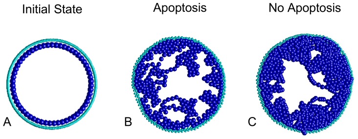
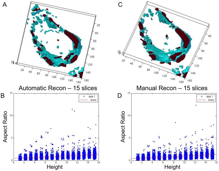
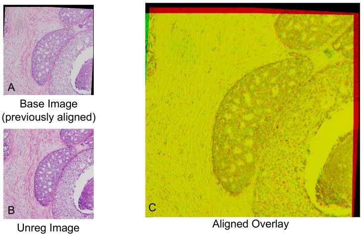
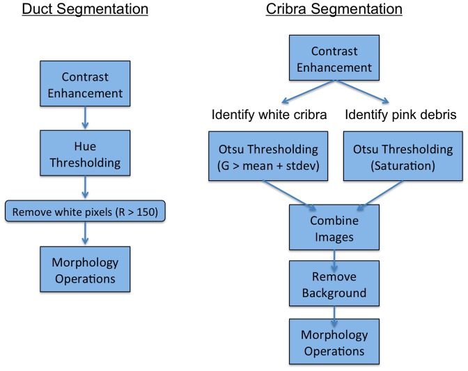
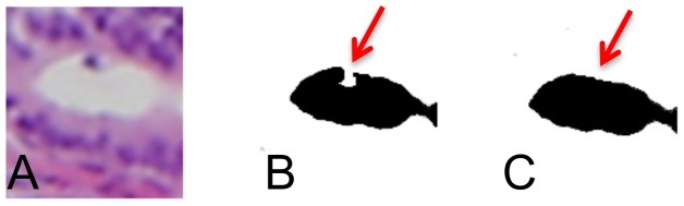
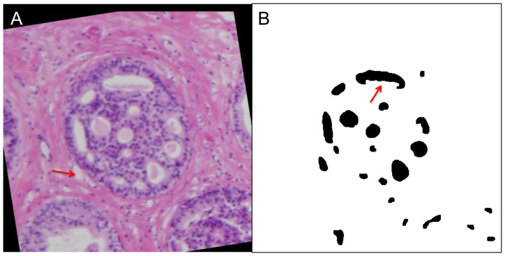
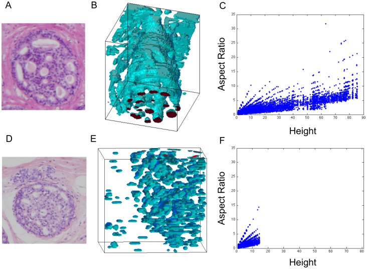
Ductal carcinoma in situ (DCIS) is a pre-invasive carcinoma of the breast that exhibits several distinct morphologies but the link between morphology and patient outcome is not clear. We hypothesize that different mechanisms of growth may still result in similar 2D morphologies, which may look different in 3D. To elucidate the connection between growth and 3D morphology, we reconstruct the 3D architecture of cribriform DCIS from resected patient material. We produce a fully automated algorithm that aligns, segments, and reconstructs 3D architectures from microscopy images of 2D serial sections from human specimens. The alignment algorithm is based on normalized cross correlation, the segmentation algorithm uses histogram equilization, Otsu's thresholding, and morphology techniques to segment the duct and cribra. The reconstruction method combines these images in 3D. We show that two distinct 3D architectures are indeed found in samples whose 2D histological sections are similarly identified as cribriform DCIS. These differences in architecture support the hypothesis that luminal spaces may form due to different mechanisms, either isolated cell death or merging fronds, leading to the different architectures. We find that out of 15 samples, 6 were found to have 'bubble-like' cribra, 6 were found to have 'tube-like' criba and 3 were 'unknown.' We propose that the 3D architectures found, 'bubbles' and 'tubes', account for some of the heterogeneity of the disease and may be prognostic indicators of different patient outcomes.
导管原位癌(DCIS)是一种乳腺的前浸润性癌,具有多种不同的形态,但形态与患者预后之间的联系尚不清楚。我们假设,不同的生长机制可能仍然导致类似的 2D 形态,而在 3D 中可能看起来不同。为了阐明生长和 3D 形态之间的联系,我们从切除的患者标本中重建筛状 DCIS 的 3D 结构。我们提出了一种全自动算法,该算法基于归一化互相关对齐、直方图均衡化、Otsu 阈值和形态学技术对来自人类标本的 2D 连续切片的显微镜图像进行分割和重建 3D 结构。该对齐算法基于归一化互相关,分割算法使用直方图均衡化、Otsu 阈值和形态学技术来分割导管和筛板。重建方法将这些图像组合在 3D 中。我们发现,在 2D 组织学切片被同样鉴定为筛状 DCIS 的样本中,确实存在两种不同的 3D 结构。这些结构上的差异支持这样的假设,即腔室可能是由于不同的机制形成的,要么是孤立的细胞死亡,要么是融合的叶,导致了不同的结构。我们发现,在 15 个样本中,有 6 个被发现具有“泡状”筛板,6 个被发现具有“管状”筛板,3 个被发现具有“未知”结构。我们提出,发现的 3D 结构,“泡状”和“管状”,解释了该疾病的一些异质性,并且可能是不同患者预后的预后指标。