P.A.I.N. Group, Center for Pain and the Brain, Children's Hospital Boston, Massachusetts General Hospital, Boston, Massachusetts, United States of America.
PLoS One. 2012;7(9):e44643. doi: 10.1371/journal.pone.0044643. Epub 2012 Sep 4.
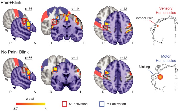
The cornea has been a focus of animal electrophysiological research for decades, but little is known regarding its cortical representation in the human brain. This study attempts to localize the somatotopic representation of the cornea to painful stimuli in human primary somatosensory cortex using functional magnetic resonance imaging (fMRI). In this case study, a subject was imaged at 3T while bright light was presented in a block-design, which either produced pain and blinking (during photophobia) or blinking alone (after recovery from photophobia). Pain and blinking produced precisely localized activations in primary somatosensory cortex and primary motor cortex. These results indicate that noxious stimulation of the cornea can produce somatotopic activation in primary somatosensory cortex. This finding opens future avenues of research to evaluate the relationship between corneal pain and central brain mechanisms relating to the development of chronic pain conditions, such as dry eye-like symptoms.
角膜一直是动物电生理学研究的焦点,但人类大脑中关于角膜的皮质代表区域却知之甚少。本研究试图使用功能磁共振成像(fMRI)定位人初级体感皮层中角膜对疼痛刺激的躯体代表区。在这项病例研究中,一名受试者在 3T 场强下进行成像,同时以块设计呈现强光,强光既产生疼痛和眨眼(在畏光时),也产生单独眨眼(在畏光恢复后)。疼痛和眨眼会在初级体感皮层和初级运动皮层产生精确的局部激活。这些结果表明,角膜的有害刺激可以在初级体感皮层中产生躯体代表区的激活。这一发现为未来的研究开辟了道路,可以评估角膜疼痛与与慢性疼痛状况(如干眼症样症状)相关的中枢大脑机制之间的关系。