• 文献检索
  • 文档翻译
  • 深度研究
  • 学术资讯
  • Suppr Zotero 插件Zotero 插件
  • 邀请有礼
  • 套餐&价格
  • 历史记录
应用&插件
Suppr Zotero 插件Zotero 插件浏览器插件Mac 客户端Windows 客户端微信小程序
定价
高级版会员购买积分包购买API积分包
服务
文献检索文档翻译深度研究API 文档MCP 服务
关于我们
关于 Suppr公司介绍联系我们用户协议隐私条款
关注我们

Suppr 超能文献

核心技术专利:CN118964589B侵权必究
粤ICP备2023148730 号-1Suppr @ 2026

文献检索

告别复杂PubMed语法,用中文像聊天一样搜索,搜遍4000万医学文献。AI智能推荐,让科研检索更轻松。

立即免费搜索

文件翻译

保留排版,准确专业,支持PDF/Word/PPT等文件格式,支持 12+语言互译。

免费翻译文档

深度研究

AI帮你快速写综述,25分钟生成高质量综述,智能提取关键信息,辅助科研写作。

立即免费体验

3D-SIM 超分辨率显微镜揭示了 FtsZ 和分裂机制的珠状排列:对触发细胞分裂的影响。

3D-SIM super resolution microscopy reveals a bead-like arrangement for FtsZ and the division machinery: implications for triggering cytokinesis.

机构信息

The ithree institute, University of Technology Sydney, Sydney, New South Wales, Australia.

出版信息

PLoS Biol. 2012;10(9):e1001389. doi: 10.1371/journal.pbio.1001389. Epub 2012 Sep 11.

DOI:10.1371/journal.pbio.1001389
PMID:22984350
原文链接:https://pmc.ncbi.nlm.nih.gov/articles/PMC3439403/
Abstract

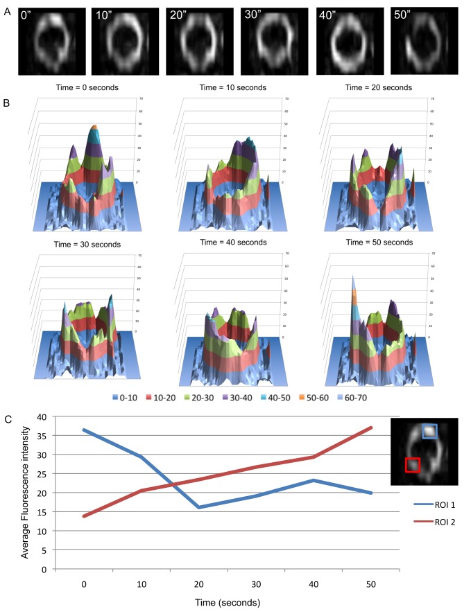
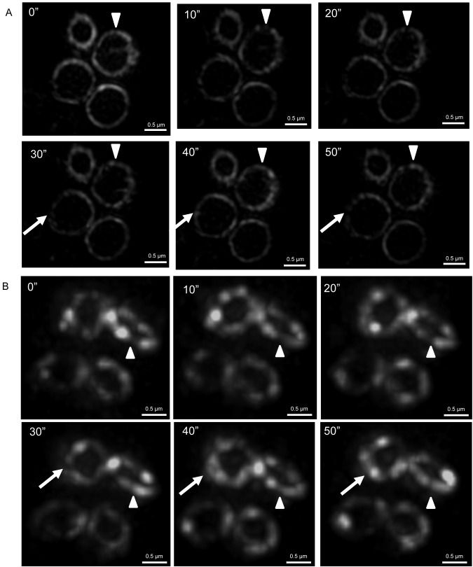
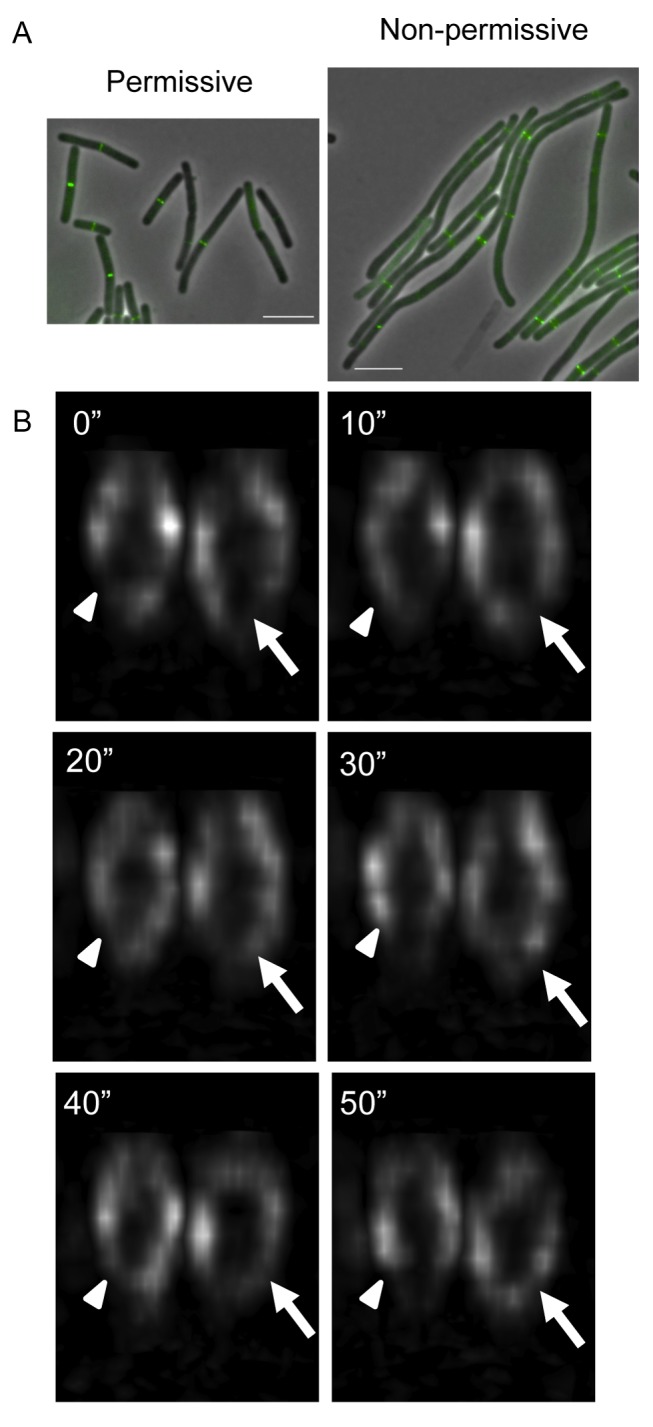
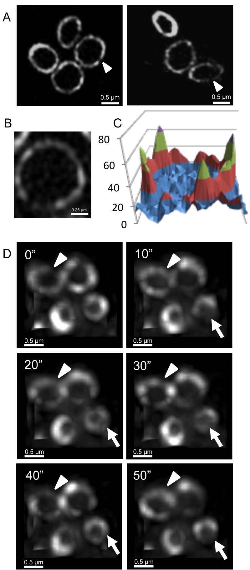
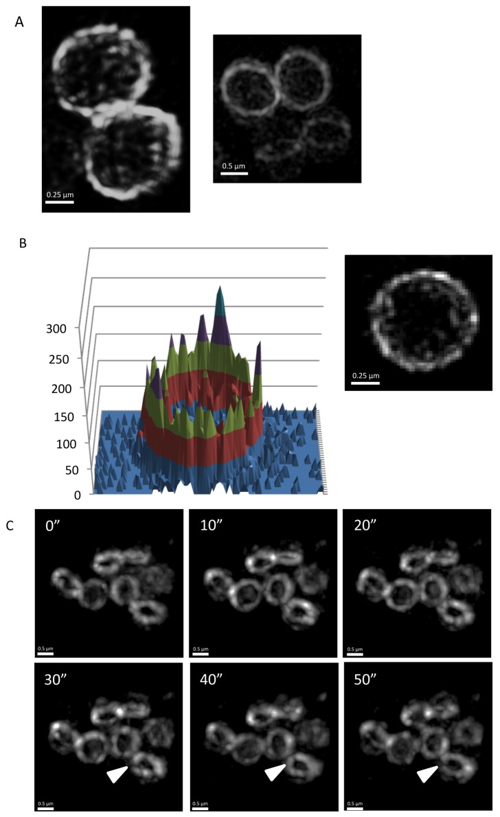
FtsZ is a tubulin-like GTPase that is the major cytoskeletal protein in bacterial cell division. It polymerizes into a ring, called the Z ring, at the division site and acts as a scaffold to recruit other division proteins to this site as well as providing a contractile force for cytokinesis. To understand how FtsZ performs these functions, the in vivo architecture of the Z ring needs to be established, as well as how this structure constricts to enable cytokinesis. Conventional wide-field fluorescence microscopy depicts the Z ring as a continuous structure of uniform density. Here we use a form of super resolution microscopy, known as 3D-structured illumination microscopy (3D-SIM), to examine the architecture of the Z ring in cells of two Gram-positive organisms that have different cell shapes: the rod-shaped Bacillus subtilis and the coccoid Staphylococcus aureus. We show that in both organisms the Z ring is composed of a heterogeneous distribution of FtsZ. In addition, gaps of fluorescence were evident, which suggest that it is a discontinuous structure. Time-lapse studies using an advanced form of fast live 3D-SIM (Blaze) support a model of FtsZ localization within the Z ring that is dynamic and remains distributed in a heterogeneous manner. However, FtsZ dynamics alone do not trigger the constriction of the Z ring to allow cytokinesis. Lastly, we visualize other components of the divisome and show that they also adopt a bead-like localization pattern at the future division site. Our data lead us to propose that FtsZ guides the divisome to adopt a similar localization pattern to ensure Z ring constriction only proceeds following the assembly of a mature divisome.

摘要

FtsZ 是一种类似于微管的 GTP 酶,是细菌细胞分裂的主要细胞骨架蛋白。它在分裂部位聚合形成一个环,称为 Z 环,并作为支架将其他分裂蛋白招募到该部位,同时为细胞分裂提供收缩力。为了了解 FtsZ 如何发挥这些功能,需要确定 Z 环的体内结构,以及该结构如何收缩以实现细胞分裂。传统的宽场荧光显微镜将 Z 环描绘为具有均匀密度的连续结构。在这里,我们使用一种称为三维结构照明显微镜(3D-SIM)的超分辨率显微镜来检查两种具有不同细胞形状的革兰氏阳性生物(棒状枯草芽孢杆菌和球形金黄色葡萄球菌)细胞中 Z 环的结构。我们表明,在这两种生物中,Z 环由 FtsZ 的不均匀分布组成。此外,荧光间隙明显,表明它是不连续的结构。使用先进形式的快速活 3D-SIM(Blaze)的时程研究支持 FtsZ 在 Z 环内定位的模型,该模型是动态的,并保持不均匀分布。然而,FtsZ 动力学本身并不会引发 Z 环的收缩以允许细胞分裂。最后,我们可视化了分裂体的其他成分,并表明它们在未来的分裂部位也采用珠状定位模式。我们的数据使我们提出 FtsZ 引导分裂体采用类似的定位模式,以确保仅在成熟分裂体组装后才进行 Z 环收缩。

https://cdn.ncbi.nlm.nih.gov/pmc/blobs/9d1c/3439403/9271d955d5f6/pbio.1001389.g009.jpg
https://cdn.ncbi.nlm.nih.gov/pmc/blobs/9d1c/3439403/07640c36a6e0/pbio.1001389.g001.jpg
https://cdn.ncbi.nlm.nih.gov/pmc/blobs/9d1c/3439403/6bef99cdd0a4/pbio.1001389.g002.jpg
https://cdn.ncbi.nlm.nih.gov/pmc/blobs/9d1c/3439403/55d1a8071557/pbio.1001389.g003.jpg
https://cdn.ncbi.nlm.nih.gov/pmc/blobs/9d1c/3439403/9fafaefe3da9/pbio.1001389.g004.jpg
https://cdn.ncbi.nlm.nih.gov/pmc/blobs/9d1c/3439403/f761d34e6e10/pbio.1001389.g005.jpg
https://cdn.ncbi.nlm.nih.gov/pmc/blobs/9d1c/3439403/bcc06c217f87/pbio.1001389.g006.jpg
https://cdn.ncbi.nlm.nih.gov/pmc/blobs/9d1c/3439403/1fcb45414293/pbio.1001389.g007.jpg
https://cdn.ncbi.nlm.nih.gov/pmc/blobs/9d1c/3439403/6147d25e20eb/pbio.1001389.g008.jpg
https://cdn.ncbi.nlm.nih.gov/pmc/blobs/9d1c/3439403/9271d955d5f6/pbio.1001389.g009.jpg

相似文献

1
3D-SIM super resolution microscopy reveals a bead-like arrangement for FtsZ and the division machinery: implications for triggering cytokinesis.3D-SIM 超分辨率显微镜揭示了 FtsZ 和分裂机制的珠状排列:对触发细胞分裂的影响。
PLoS Biol. 2012;10(9):e1001389. doi: 10.1371/journal.pbio.1001389. Epub 2012 Sep 11.
2
A new assembly pathway for the cytokinetic Z ring from a dynamic helical structure in vegetatively growing cells of Bacillus subtilis.枯草芽孢杆菌营养生长细胞中细胞分裂Z环从动态螺旋结构形成的新组装途径。
Mol Microbiol. 2007 Apr;64(2):487-99. doi: 10.1111/j.1365-2958.2007.05673.x.
3
Single-molecule imaging reveals that Z-ring condensation is essential for cell division in Bacillus subtilis.单分子成像揭示了 Z 环凝聚对于枯草芽孢杆菌细胞分裂的重要性。
Nat Microbiol. 2021 May;6(5):553-562. doi: 10.1038/s41564-021-00878-z. Epub 2021 Mar 18.
4
Super-resolution imaging of the bacterial cytokinetic protein FtsZ.细菌细胞分裂蛋白FtsZ的超分辨率成像
Micron. 2011 Jun;42(4):336-41. doi: 10.1016/j.micron.2010.09.003. Epub 2010 Sep 15.
5
Bacteriophage SP01 Gene Product 56 Inhibits Bacillus subtilis Cell Division by Interacting with FtsL and Disrupting Pbp2B and FtsW Recruitment.噬菌体 SP01 基因产物 56 通过与 FtsL 相互作用并破坏 Pbp2B 和 FtsW 的招募来抑制枯草芽孢杆菌的细胞分裂。
J Bacteriol. 2020 Dec 18;203(2). doi: 10.1128/JB.00463-20.
6
Peptidoglycan synthesis drives an FtsZ-treadmilling-independent step of cytokinesis.肽聚糖合成驱动了胞质分裂的一个 FtsZ treadmilling 非依赖的步骤。
Nature. 2018 Feb 22;554(7693):528-532. doi: 10.1038/nature25506. Epub 2018 Feb 14.
7
An essential cell division protein directly regulates FtsZ dynamics.一种重要的细胞分裂蛋白直接调控 FtsZ 的动态变化。
Elife. 2018 Oct 2;7:e38856. doi: 10.7554/eLife.38856.
8
Progression of the late-stage divisome is unaffected by the depletion of the cytoplasmic FtsZ pool.后期分裂体的进展不受细胞质 FtsZ 池耗尽的影响。
Commun Biol. 2021 Mar 1;4(1):270. doi: 10.1038/s42003-021-01789-9.
9
Noc Corrals Migration of FtsZ Protofilaments during Cytokinesis in Bacillus subtilis.Noc 阻止枯草芽孢杆菌细胞分裂过程中 FtsZ 原丝的迁移。
mBio. 2021 Feb 2;12(1):e02964-20. doi: 10.1128/mBio.02964-20.
10
3D-SIM super-resolution of FtsZ and its membrane tethers in Escherichia coli cells.大肠杆菌细胞中FtsZ及其膜系链蛋白的3D-SIM超分辨率成像
Biophys J. 2014 Oct 21;107(8):L17-L20. doi: 10.1016/j.bpj.2014.08.024.

引用本文的文献

1
EzrA promotes Z-ring formation through interaction of its QNR motif with FtsA.EzrA通过其QNR基序与FtsA的相互作用促进Z环的形成。
J Bacteriol. 2025 Jul 24;207(7):e0012525. doi: 10.1128/jb.00125-25. Epub 2025 Jul 3.
2
A FtsZ cis disassembly element acts in Z-ring assembly during bacterial cell division.一个FtsZ顺式解聚元件在细菌细胞分裂过程中的Z环组装中起作用。
Nat Commun. 2025 Jun 4;16(1):5194. doi: 10.1038/s41467-025-60517-7.
3
Application of nanotags and nanobodies for live cell single-molecule imaging of the Z-ring in Escherichia coli.

本文引用的文献

1
EzrA contributes to the regulation of cell size in Staphylococcus aureus.EzrA 参与调控金黄色葡萄球菌的细胞大小。
PLoS One. 2011;6(11):e27542. doi: 10.1371/journal.pone.0027542. Epub 2011 Nov 14.
2
Inside-out Z rings--constriction with and without GTP hydrolysis.内外翻转 Z 环——有无 GTP 水解的收缩。
Mol Microbiol. 2011 Jul;81(2):571-9. doi: 10.1111/j.1365-2958.2011.07716.x. Epub 2011 Jun 16.
3
Multiple essential roles for EzrA in cell division of Staphylococcus aureus.EzrA 在金黄色葡萄球菌细胞分裂中的多重必需作用。
纳米标签和纳米抗体在活细胞中单分子成像大肠杆菌 Z 环中的应用。
Curr Genet. 2023 Jun;69(2-3):153-163. doi: 10.1007/s00294-023-01266-2. Epub 2023 Apr 6.
4
Microfluidic Organ-on-A-chip: A Guide to Biomaterial Choice and Fabrication.微流控器官芯片:生物材料选择与制造指南。
Int J Mol Sci. 2023 Feb 6;24(4):3232. doi: 10.3390/ijms24043232.
5
Three-dimensional structured illumination microscopy with enhanced axial resolution.三维结构光照明显微镜,轴向分辨率增强。
Nat Biotechnol. 2023 Sep;41(9):1307-1319. doi: 10.1038/s41587-022-01651-1. Epub 2023 Jan 26.
6
FtsN maintains active septal cell wall synthesis by forming a processive complex with the septum-specific peptidoglycan synthases in E. coli.FtsN 通过与大肠杆菌中特定于隔膜的肽聚糖合成酶形成一个连续的复合物来维持活跃的隔膜细胞壁合成。
Nat Commun. 2022 Sep 30;13(1):5751. doi: 10.1038/s41467-022-33404-8.
7
[Comparison and progress review of various super-resolution fluorescence imaging techniques].[各种超分辨率荧光成像技术的比较与进展综述]
Se Pu. 2021 Oct;39(10):1055-1064. doi: 10.3724/SP.J.1123.2021.06015.
8
The Division Defect of a Double Mutant Can Be Suppressed by Spx-Dependent and Spx-Independent Mechanisms.双突变体的分裂缺陷可以通过 Spx 依赖和不依赖的机制得到抑制。
J Bacteriol. 2021 Aug 20;203(18):e0024921. doi: 10.1128/JB.00249-21.
9
Viperin interacts with PEX19 to mediate peroxisomal augmentation of the innate antiviral response.Viperin 通过与 PEX19 相互作用来介导先天抗病毒反应的过氧化物酶体增强。
Life Sci Alliance. 2021 Jun 9;4(7). doi: 10.26508/lsa.202000915. Print 2021 Jul.
10
Asymmetric localization of the cell division machinery during sporulation.细胞分裂机制在孢子形成过程中的不对称定位。
Elife. 2021 May 21;10:e62204. doi: 10.7554/eLife.62204.
Mol Microbiol. 2011 Apr;80(2):542-55. doi: 10.1111/j.1365-2958.2011.07591.x. Epub 2011 Mar 14.
4
FtsZ in bacterial cytokinesis: cytoskeleton and force generator all in one.细菌胞质分裂中的 FtsZ:细胞骨架和力发生器合二为一。
Microbiol Mol Biol Rev. 2010 Dec;74(4):504-28. doi: 10.1128/MMBR.00021-10.
5
A simple plasmid-based system that allows rapid generation of tightly controlled gene expression in Staphylococcus aureus.一种简单的基于质粒的系统,可在金黄色葡萄球菌中快速产生严格控制的基因表达。
Microbiology (Reading). 2011 Mar;157(Pt 3):666-676. doi: 10.1099/mic.0.045146-0. Epub 2010 Nov 25.
6
Super-resolution imaging of the bacterial cytokinetic protein FtsZ.细菌细胞分裂蛋白FtsZ的超分辨率成像
Micron. 2011 Jun;42(4):336-41. doi: 10.1016/j.micron.2010.09.003. Epub 2010 Sep 15.
7
In vivo structure of the E. coli FtsZ-ring revealed by photoactivated localization microscopy (PALM).活细胞中由光激活定位显微镜(PALM)揭示的大肠杆菌 FtsZ 环的结构。
PLoS One. 2010 Sep 13;5(9):e12682. doi: 10.1371/journal.pone.0012680.
8
Fast live simultaneous multiwavelength four-dimensional optical microscopy.快速实时同步多波长四维光学显微镜。
Proc Natl Acad Sci U S A. 2010 Sep 14;107(37):16016-22. doi: 10.1073/pnas.1004037107. Epub 2010 Aug 12.
9
A guide to super-resolution fluorescence microscopy.超分辨率荧光显微镜指南。
J Cell Biol. 2010 Jul 26;190(2):165-75. doi: 10.1083/jcb.201002018. Epub 2010 Jul 19.
10
FtsZ inhibition: a promising approach for antistaphylococcal therapy.FtsZ抑制:一种有前景的抗葡萄球菌治疗方法。
Drug News Perspect. 2010 Jun;23(5):295-304. doi: 10.1358/dnp.2010.23.5.1429489.