Unidade de Investigação em Eco-Etologia, Instituto Superior de Psicologia Aplicada, Lisboa, Portugal.
PLoS One. 2012;7(9):e44086. doi: 10.1371/journal.pone.0044086. Epub 2012 Sep 11.
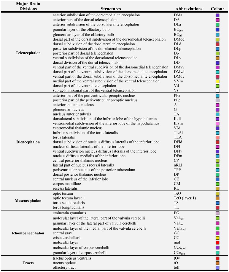
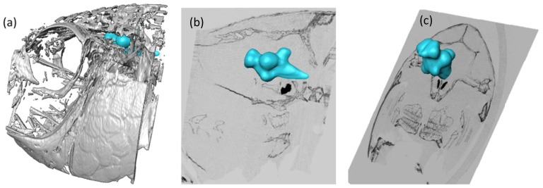
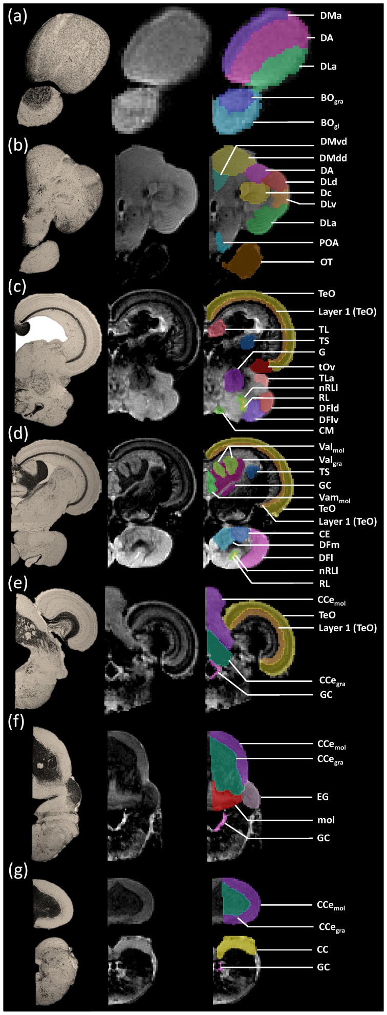
The African cichlid Oreochromis mossambicus (Mozambique tilapia) has been used as a model system in a wide range of behavioural and neurobiological studies. The increasing number of genetic tools available for this species, together with the emerging interest in its use for neurobiological studies, increased the need for an accurate hodological mapping of the tilapia brain to supplement the available histological data. The goal of our study was to elaborate a three-dimensional, high-resolution digital atlas using magnetic resonance imaging, supported by Nissl staining. Resulting images were viewed and analysed in all orientations (transverse, sagittal, and horizontal) and manually labelled to reveal structures in the olfactory bulb, telencephalon, diencephalon, optic tectum, and cerebellum. This high resolution tilapia brain atlas is expected to become a very useful tool for neuroscientists using this fish model and will certainly expand their use in future studies regarding the central nervous system.
非洲慈鲷 Oreochromis mossambicus(莫桑比克口孵非鲫)已被广泛应用于行为学和神经生物学研究中。由于该物种有越来越多的遗传工具可用,同时人们对其在神经生物学研究中的应用也越来越感兴趣,因此需要对罗非鱼大脑进行精确的神经解剖学图谱绘制,以补充现有的组织学数据。我们的研究目标是使用磁共振成像(MRI)结合尼氏染色制作一个三维、高分辨率的数字图谱。结果图像以所有方向(横切面、矢状面和水平面)进行查看和分析,并进行手动标记以显示嗅球、端脑、间脑、视顶盖和小脑的结构。这个高分辨率的罗非鱼脑图谱有望成为使用这种鱼类模型的神经科学家的一个非常有用的工具,并将在未来关于中枢神经系统的研究中进一步扩大其应用。