Department of Radiology, University of Wisconsin, Madison, WI, USA.
J Nucl Med. 2012 Nov;53(11):1748-54. doi: 10.2967/jnumed.112.105460. Epub 2012 Sep 17.
Upregulation of tissue factor (TF) expression leads to increased patient morbidity and mortality in many solid tumor types. The goal of this study was to develop a PET tracer for imaging of TF expression in pancreatic cancer.
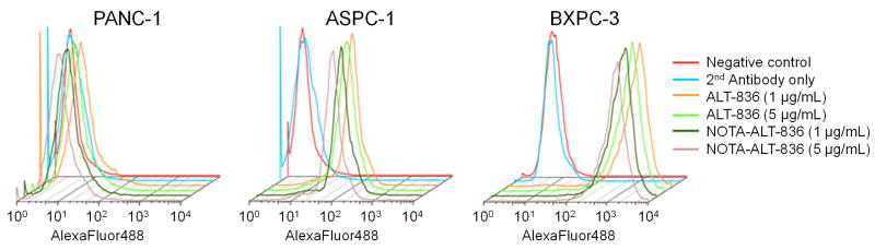
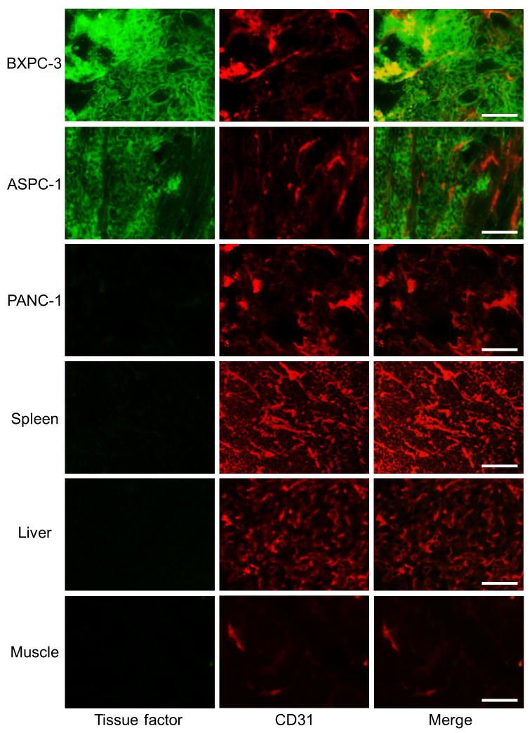
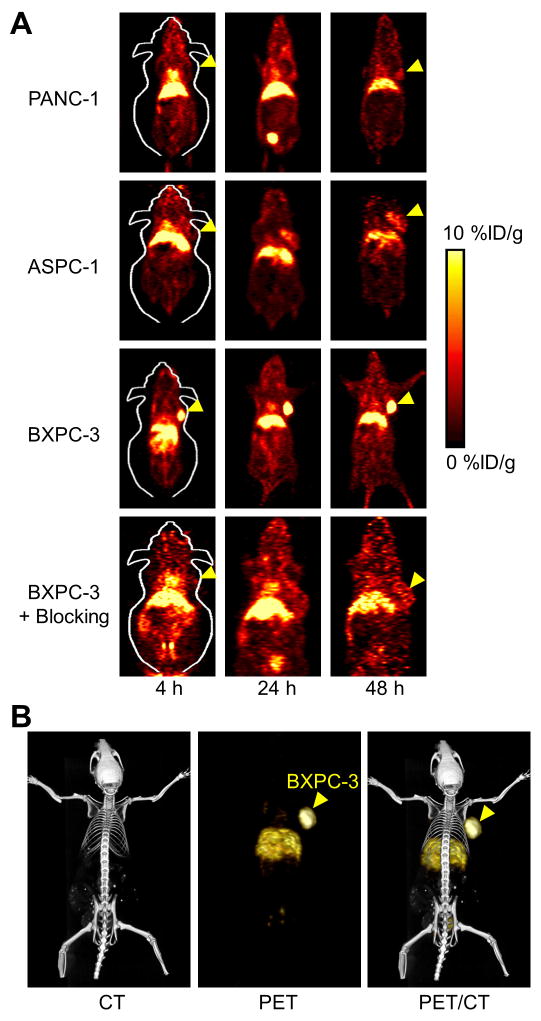
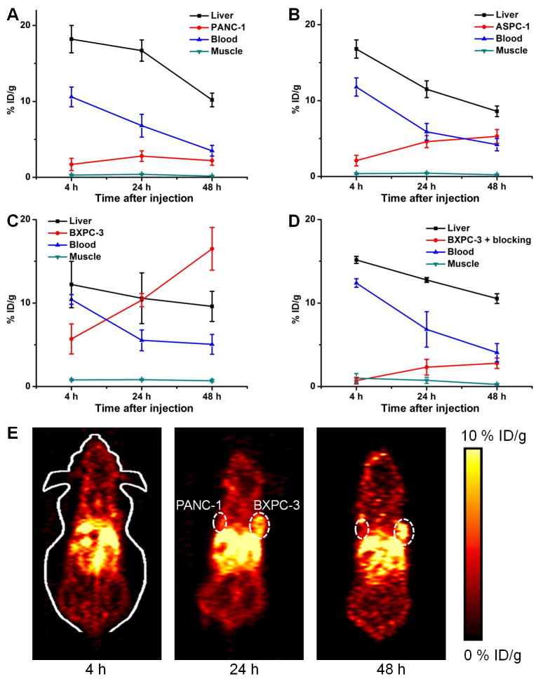
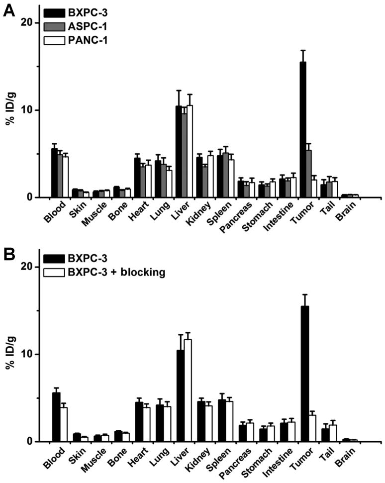
ALT-836, a chimeric antihuman TF monoclonal antibody, was conjugated to 2-S-(4-isothiocyanatobenzyl)-1,4,7-triazacyclononane-1,4,7-triacetic acid (p-SCN-Bn-NOTA) and labeled with (64)Cu. To compare the TF binding affinity of ALT-836 and NOTA-ALT-836, flow cytometry analysis was performed in 3 pancreatic cancer cell lines with different expression levels of TF (from low to high: PANC-1, ASPC-1, and BXPC-3). PET, biodistribution, blocking, and histology studies were performed on pancreatic tumor-bearing mice to evaluate the ability and specificity of (64)Cu-NOTA-ALT-836 to target TF in vivo.
There was no difference in TF binding affinity between ALT-836 and NOTA-ALT-836. (64)Cu-labeling was achieved with high yield and specific activity. Serial PET revealed that the uptake of (64)Cu-NOTA-ALT-836 in BXPC-3 tumors (high TF expression) was 5.7 ± 1.8, 10.4 ± 0.8, and 16.5 ± 2.6 percentage injected dose per gram at 4, 24, and 48 h after injection, respectively (n = 4), significantly higher than that in the PANC-1 and ASPC-1 tumors. Biodistribution data as measured by γ-counting were consistent with the PET findings. Blocking experiments and histology further confirmed the TF specificity of (64)Cu-NOTA-ALT-836.
Herein we report the first successful PET imaging of TF expression. Persistent and TF-specific uptake of (64)Cu-NOTA-ALT-836 was observed in pancreatic cancer models.
开发用于成像胰腺癌组织因子(TF)表达的 PET 示踪剂。
将嵌合抗人 TF 单克隆抗体 ALT-836 与 2-S-(4-异硫氰酸苄基)-1,4,7-三氮杂环壬烷-1,4,7-三乙酸(p-SCN-Bn-NOTA)缀合,并标记(64)Cu。为了比较 ALT-836 和 NOTA-ALT-836 的 TF 结合亲和力,在 TF 表达水平不同(从低到高:PANC-1、ASPC-1 和 BXPC-3)的 3 种胰腺癌细胞系中进行了流式细胞术分析。在荷胰腺肿瘤小鼠中进行了 PET、生物分布、阻断和组织学研究,以评估(64)Cu-NOTA-ALT-836 在体内靶向 TF 的能力和特异性。
ALT-836 和 NOTA-ALT-836 之间没有 TF 结合亲和力的差异。(64)Cu 标记具有高产率和比活性。连续 PET 显示,(64)Cu-NOTA-ALT-836 在 BXPC-3 肿瘤(高 TF 表达)中的摄取率分别为 4、24 和 48 h 后注射后每克 5.7 ± 1.8、10.4 ± 0.8 和 16.5 ± 2.6% 注入剂量(n = 4),显著高于 PANC-1 和 ASPC-1 肿瘤。γ计数测量的生物分布数据与 PET 结果一致。阻断实验和组织学进一步证实了(64)Cu-NOTA-ALT-836 的 TF 特异性。
本文首次成功进行了 TF 表达的 PET 成像。在胰腺癌细胞模型中观察到(64)Cu-NOTA-ALT-836 的持续且 TF 特异性摄取。