Department of Anesthesia and Perioperative Care, University of California, San Francisco, San Francisco, California 94143-0542, USA.
Anesthesiology. 2012 Nov;117(5):1080-90. doi: 10.1097/ALN.0b013e31826f8d86.
Propofol in the early postnatal period has been shown to cause brain cell death. One proposed mechanism for cognitive dysfunction after anesthesia is alteration of neural stem cell function and neurogenesis. We examined the effect of propofol on neural precursor or stem cells (NPCs) grown in vitro.
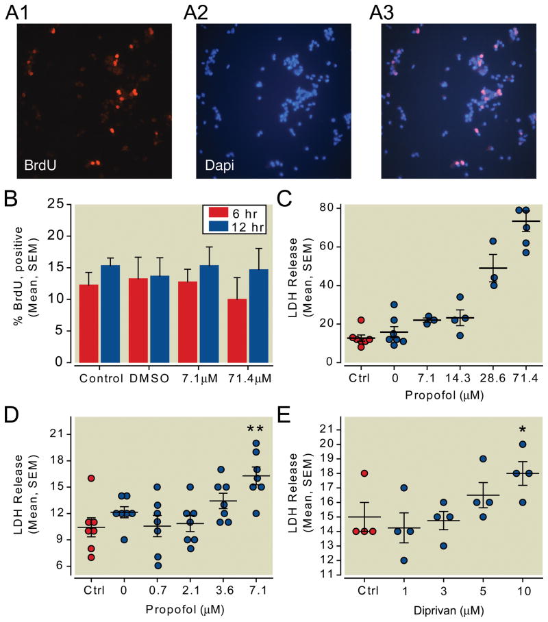
Hippocampal-derived NPCs from postnatal day 2 rats were exposed to propofol or Diprivan. NPCs were then analyzed for bromodeoxyuridine incorporation to measure proliferation. Cell death was measured by lactate dehydrogenase release. Immunocytochemistry was used to evaluate the expression of neuronal and glial markers in differentiating NPCs exposed to propofol.
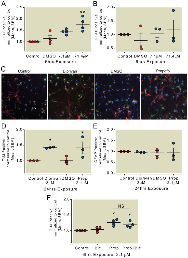
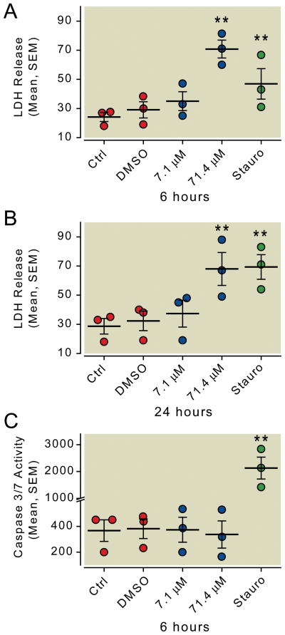
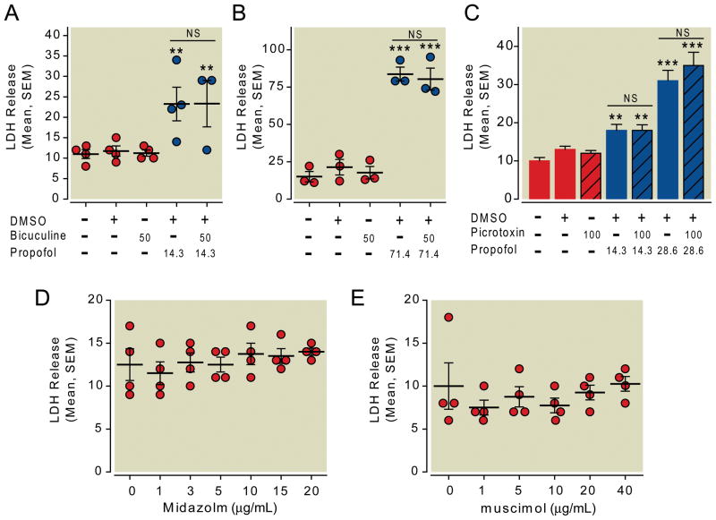
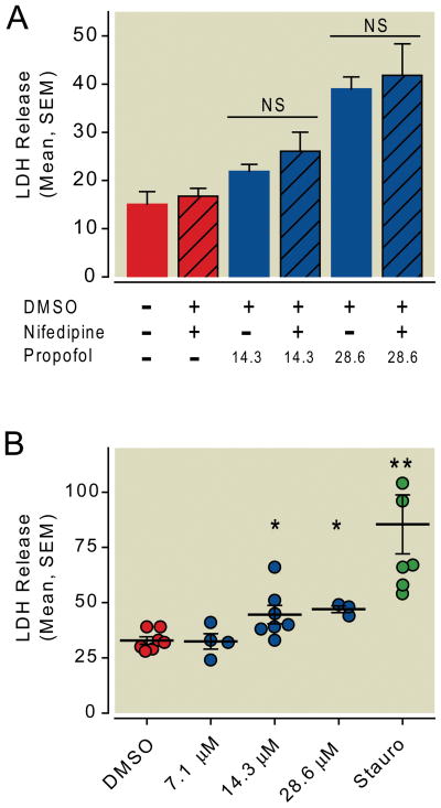
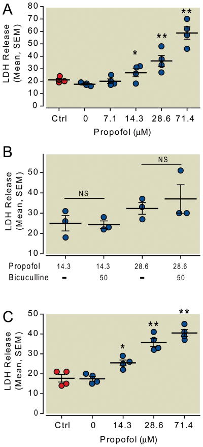
Propofol dose dependently increases the release of lactate dehydrogenase from NPCs under both proliferating and differentiating conditions at supraclinical concentrations (more than 7.1 µM). Both Diprivan and propofol had the same effect on NPCs. Propofol-mediated release of lactate dehydrogenase is not inhibited by blocking the γ-aminobutyric acid type A receptor or extracellular calcium influx and is not mediated by caspase-3/7. Direct γ-aminobutyric acid type A receptor activation did not have the same effect. In differentiating NPCs, 6 h of propofol at 2.1 µM increased the number neurons but not glial cells 4 days later. Increased neuronal differentiation was not blocked by bicuculline.
Only supraclinical concentrations of propofol or Diprivan kill NPCs in culture by a non-γ-aminobutyric acid type A, noncaspase-3 mechanism. Clinically relevant doses of propofol increase neuronal fate choice by a non-γ-aminobutyric acid type A mechanism.
研究表明,新生期的丙泊酚会导致脑细胞死亡。麻醉后认知功能障碍的一个提出的机制是神经干细胞功能和神经发生的改变。我们研究了丙泊酚对体外培养的神经前体细胞或干细胞(NPCs)的影响。
从出生后第 2 天的大鼠海马中分离 NPCs 并暴露于丙泊酚或异丙酚。然后通过溴脱氧尿苷掺入来测量增殖来分析 NPCs 的增殖情况。通过乳酸脱氢酶释放来测量细胞死亡。用免疫细胞化学检测暴露于丙泊酚的分化 NPCs 中神经元和神经胶质标记物的表达。
在超临床浓度(超过 7.1 µM)下,丙泊酚在增殖和分化条件下均呈剂量依赖性地增加 NPCs 中乳酸脱氢酶的释放。异丙酚和丙泊酚对 NPCs 有相同的作用。丙泊酚介导的乳酸脱氢酶释放不受阻断γ-氨基丁酸 A 型受体或细胞外钙内流的抑制,也不由 caspase-3/7 介导。直接激活γ-氨基丁酸 A 型受体没有相同的效果。在分化的 NPCs 中,2.1 µM 的丙泊酚作用 6 小时后,4 天后增加神经元的数量,但不增加神经胶质细胞的数量。增加的神经元分化不能被荷包牡丹碱阻断。
只有超临床浓度的丙泊酚或异丙酚通过非γ-氨基丁酸 A、非 caspase-3 机制杀死培养中的 NPCs。临床相关剂量的丙泊酚通过非γ-氨基丁酸 A 机制增加神经元命运选择。